3T Insula MRI Anatomy Ultra-high-resolution 1.5mm Slices (Subscription-Based Premium Access)
Explore our advanced insula MRI anatomy library, featuring high-resolution labelled MRI images of the insular cortex and surrounding deep brain structures in axial and sagittal planes. This module is designed to help users understand the complex anatomy of the insula, opercula, basal ganglia, and adjacent white matter pathways.
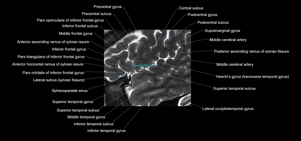
The insula anatomy section includes the anterior and posterior insular cortex, short and long insular gyri, central insular sulcus, circular sulcus, frontal operculum, parietal operculum, temporal operculum, extreme capsule, claustrum, external capsule, putamen, internal capsule, and middle cerebral artery branches. These structures are important in stroke imaging, epilepsy, tumour localisation, neurosurgical planning, and advanced neuroanatomy.
All MRI images are manually labelled slice by slice in a textbook-style format. The labelling is performed without AI auto-labelling tools, allowing careful identification of subtle structures and accurate anatomical relationships. This makes the module valuable for radiologists, neurosurgeons, neurologists, radiology students, reporting radiographers, and anyone learning detailed brain anatomy.
The interactive scroller allows users to move through the insular region smoothly, helping them understand how the insula appears across different planes and levels. The viewer is optimised for desktop and mobile use and includes a full-screen option for focused study.
The bottom toolbar allows users to switch between axial and sagittal planes. Voice and text search make it easy to locate key structures and access detailed descriptions of their appearance and anatomical relationships.
The 10x zoom function allows close inspection of the insular cortex, opercula, middle cerebral artery branches, and adjacent white matter pathways without loss of image quality.
The insula MRI anatomy module includes dynamic Level 1 and Level 2 quizzes, helping users revise anatomy actively and build long-term understanding of this complex deep cortical region.
MRI insula Axial 1.5 mm Deep Resolve Labelled Image(sample)
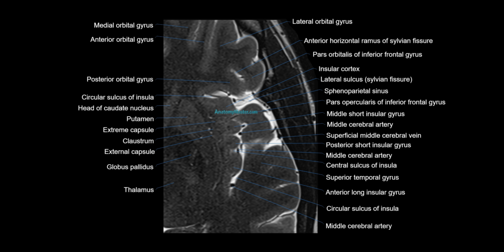
MRI insula sagittal 1.5 mm Deep Resolve Labelled Image(sample)