MRI VIBE(LAVA-XV/THRIVE/TIGRE) Post Contrast
MRI image appearance VIBE Post Contrast
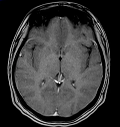
The easiest way to identify T1 VIBE post-gadolinium images is to look for blood vessels in the body (e.g., arteries and veins in the brain, neck, chest, abdomen, upper limbs, and lower limbs). Blood vessels and pathologies with high vascularity appear bright in T1 VIBE post-gadolinium images. All the other characteristics of the T1 VIBE post-gadolinium images remain the same as the T1 VIBE images.
Tissues and their T1 VIBE post gadolinium appearance
Fluids : – dark (Ureters and bladder appear bright due to the contrast excretion)
Bone marrow : – equal to or higher than that of muscle (fatty marrow is usually bright)
Muscles- gray
Kidneys :- bright
Liver :-bright
Spleen :- bright
Pancreas :- bright
Bowel walls :- bright
Prostate :- bright
Uterus:- bright
Moving blood : – bright
White matter : – whiter
Gray matter : – gray
Bone : – dark
Fat : – bright
Air : – dark
Use
- Useful for abdominal imaging
- Useful for prostate imaging
- Useful for brain imaging
Pathological appearance
Pathologies with hypervascularization will appear bright on T1 VIBE post-contrast gadolinium images. Examples of such pathologies include tumors like hemangioma, lymphangioma, hemangioendothelioma, Kaposi’s sarcoma, angiosarcoma, hemangioblastoma, as well as inflammatory processes like discitis, meningitis, synovitis, arthritis, and osteomyelitis. Pathological processes with no vascularity will remain unenhanced, appearing dark on T1 VIBE post-contrast gadolinium images.
VIBE AXIAL POST CONTRAST SEQUENCE USED IN PROSTATE IMAGING

VIBE AXIAL POST CONTRAST SEQUENCE USED IN UNCOOPERATIVE PATIENTS BRAIN IMAGING