FLASH(SPGR/T1-FFE/RF Spoiled SARGE/RSSG/FAstFE/S Tage/T1-FAST) Fat Sat Post GD
MRI image appearance
The easiest way to identify T1 FLASH fat saturated post gadolinium images is to look for adipose tissues (e.g. subcutaneous fat and fat in bone marrow) and blood vessels in the body (e.g arteries and veins in chest, abdomen, upper limbs and lower limbs). Areas contain adipose tissues appear as dark on T1 FLASH fat saturated post gadolinium images. Blood vessels and pathologies with high vascularity appear as bright on T1 FLASH fat saturated post gadolinium images. All the other characteristics of the T1 FLASH fat saturated post gadolinium images remain same as the T1 FLASH image.
Tissues and their T1 FLASH fat saturated post gadolinium appearance
Fluids : – dark (Ureters and bladder appear as bright due to the contrast excretion)
Bone marrow : – dark
Muscles- gray
Kidneys :- bright
Liver :-bright
Spleen :- bright
Pancreas :- bright
Bowel walls :- bright
Prostate :- bright
Uterus:- bright
Moving blood : – bright
White matter : – whiter
Gray matter : – gray
Bone : – dark
Fat : – dark
Air : – dark
Use
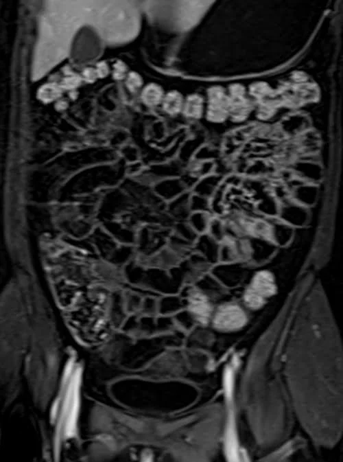
- Very useful for small bowel imaging
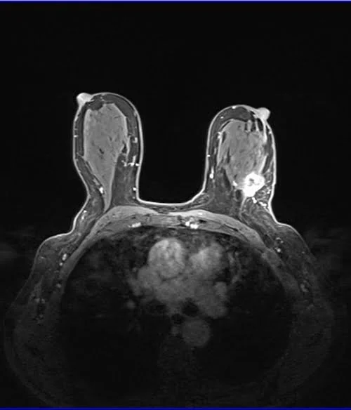
- Very useful for breast imaging
- Very useful for pancreas imaging
- Very useful for kidney imaging
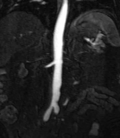
- Very useful for angiography imaging
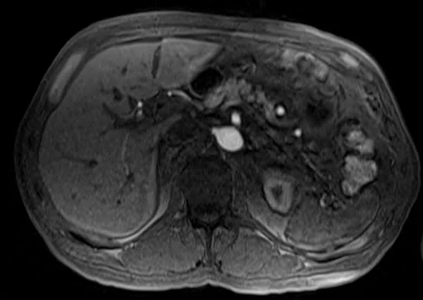
- Very useful for liver imaging
- Very useful for kidneys imaging
- Very useful for urography imaging
- Useful for chest imaging
- Useful for abdominal imaging
- Useful dynamic pelvis imaging
- Useful for extremity 3D imaging
Pathological appearance
Pathologies with adipose tissues content will appear as dark on T1 FLASH fat saturated post gadolinium images (e.g. lipoma). Pathologies with hypervascularization will appear as bright on T1 FLASH fat saturated post gadolinium images (e.g. tumours like hemangioma, Lymphangioma, hemangioendothelioma, Kaposi sarcoma, angiosarcoma, hemangioblastoma etc. and inflammatory processes like discitis, meningitis, synovitis, arthritis, osteomyelitis etc.). Pathological processes with no vascularity will remain unenhanced (appear dark in a T1 FLASH fat saturated post gadolinium image).
FLASH FAT SAT AXIAL POST CONTRAST SEQUENCE USED IN BREAST IMAGING
FLASH FAT SAT CORONAL POST CONTRAST SEQUENCE USED IN RENAL ANGIOGRAPHY IMAGING
FLASH FAT SAT POST CONTRAST AXIAL SEQUENCE USED IN LIVER IMAGING
FLASH FAT SAT CORONAL POST CONTRAST SEQUENCE USED IN SMALL BOWEL IMAGING