Phase-sensitive inversion recovery (PSIR) MRI
Phase-sensitive inversion recovery (PSIR) is a specialized technique used in magnetic resonance imaging (MRI) that enhances image quality by improving the contrast of the images. It’s particularly useful in cases where precise differentiation of tissues is required, such as in cardiac imaging or in the detection of lesions in the brain.
Physics of Phase-sensitive inversion recovery MRI
- Inversion Recovery (IR): PSIR starts with a standard inversion recovery sequence. This involves applying an initial pulse to invert the magnetization of tissues (flipping it by 180 degrees). This is followed by a period of time, known as the inversion time (TI), during which different tissues recover their magnetization at different rates.
- Phase Sensitivity: Traditional IR sequences use magnitude reconstruction, which only considers the magnitude of the signal, ignoring its phase. PSIR, however, uses the phase information as well as the magnitude. This makes it possible to detect subtle differences in signal that might be lost in standard IR techniques.
- Improved Contrast and Detection: By using both the phase and magnitude information, PSIR provides better contrast between different tissues. This is particularly effective for detecting small differences in tissue properties, such as between scar tissue and healthy tissue in the heart, or different types of brain tissue.
Applications of PSIR MRI Sequence
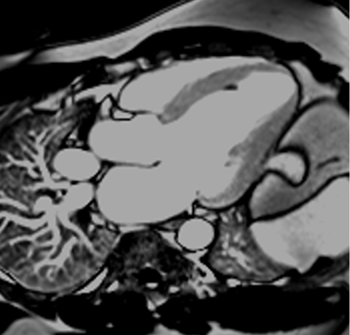
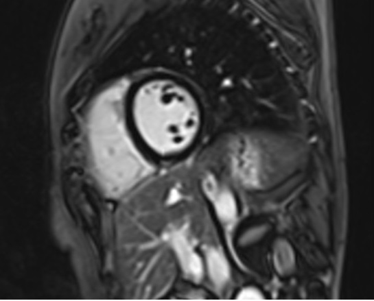
Cardiac MRI: Immediate Post-Contrast
Shortly after the administration of contrast agents, PSIR can be used to enhance the visualization of vascular structures and myocardial perfusion. The immediate phase primarily benefits from the high contrast provided by gadolinium-based contrast agents circulating in the bloodstream. Immediate post-contrast PSIR sequences are mainly used for the visualization of clots and tumors.
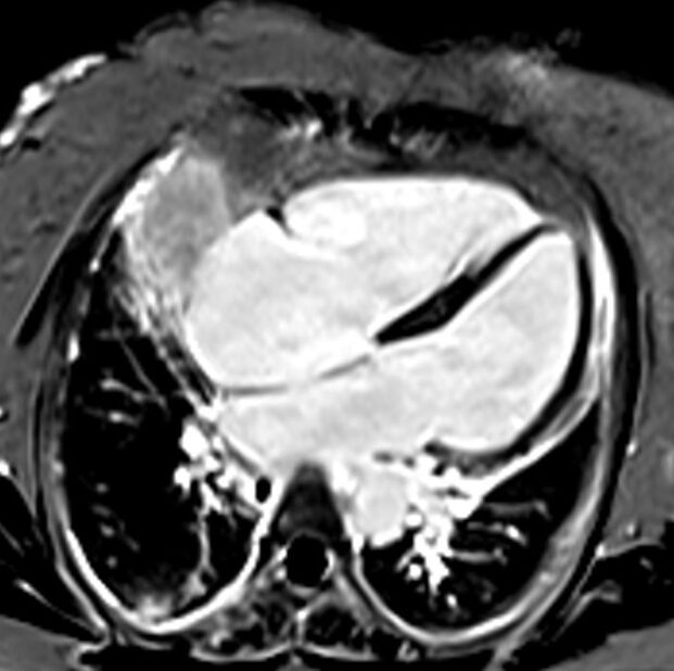
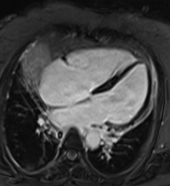
Phase-contrast (PC) 2-Chambar sequence with TI 450 used in Immediate Post-Contrast imaging
Phase sensitive IR Image
Magnitude IR Image

Cardiac MRI: Delayed Post-Contrast
In delayed post-contrast imaging, also known as late gadolinium enhancement (LGE), PSIR is particularly useful. Here, the inversion time (TI) is critically selected to nullify the signal from the normal myocardium. This TI is often determined by a Look-Locker sequence . By setting the TI such that the normal myocardium’s signal is zero (nulled), any remaining enhanced regions (indicative of fibrosis or scar tissue) are conspicuously bright against the dark background of the healthy myocardial tissue.
The ability of PSIR to utilize phase information ensures that even if there is slight variability in the actual TI used (due to patient-specific or timing variations), the contrast and visibility of pathological tissues are maintained, reducing the likelihood of misinterpretation.
Phase-contrast (PC) 4-Chambar sequence with TI 245 used in Delayed post-contrast imaging
Phase sensitive IR Image
Phase sensitive IR Image
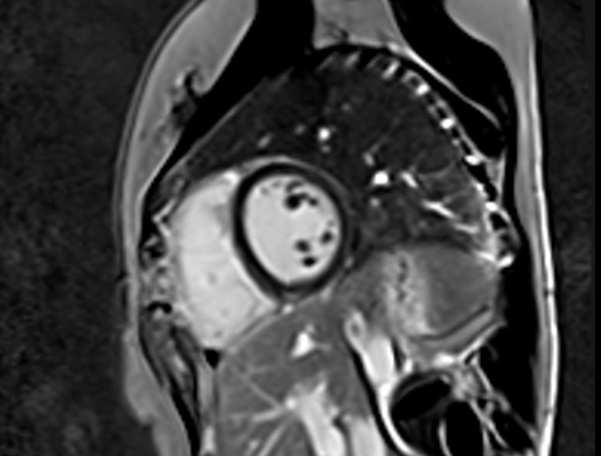
Phase-contrast (PC) short axis sequence with TI 245 used in Delayed post-contrast imaging
Phase sensitive IR Image
Phase sensitive IR Image
Neuroimaging
PSIR is extensively used to enhance the contrast in imaging of brain tissues, particularly in detecting lesions or abnormalities in multiple sclerosis.
References
- Kellman, P., Arai, A. E., McVeigh, E. R., & Aletras, A. H. (2002). Phase-Sensitive Inversion Recovery for Detecting Myocardial Infarction Using Gadolinium-Delayed Hyperenhancement. Magn Reson Med, 47(2), 372–383. https://doi.org/10.1002/mrm.10051
- Ginami, G., Neji, R., Rashid, I., Chiribiri, A., Ismail, T. F., Botnar, R. M., & Prieto, C. (2017). 3D whole-heart phase sensitive inversion recovery CMR for simultaneous black-blood late gadolinium enhancement and bright-blood coronary CMR angiography. Journal of Cardiovascular Magnetic Resonance, 19, 94.
- Favaretto, A., Poggiali, D., Lazzarotto, A., Rolma, G., Causin, F., & Gallo, P. (2015). The Parallel Analysis of Phase Sensitive Inversion Recovery (PSIR) and Double Inversion Recovery (DIR) Images Significantly Improves the Detection of Cortical Lesions in Multiple Sclerosis (MS) since Clinical Onset. PLoS One, 10(5), e0127805. https://doi.org/10.1371/journal.pone.0127805