MRI Oversampling \ Fold-Over Suppression
Oversampling
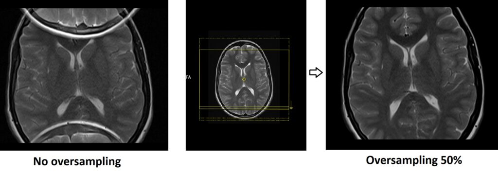
Oversampling, also referred to as “fold-over suppression” or “fold-over artifact suppression,” is a technique used in magnetic resonance imaging (MRI) to mitigate a common artifact that arises when imaging an object or region that is larger than the field of view (FOV) of the MRI scanner.
When the object being imaged extends beyond the FOV, a fold-over artifact can occur. This artifact leads to signal from one part of the object erroneously “folding over” into another part of the image, causing distortion and confusion in the resulting MRI image.

Oversampling involves acquiring additional data beyond the boundaries of the original FOV. By capturing data from areas adjacent to the FOV, the scanner can effectively capture the information needed to correct or suppress the fold-over artifact. This extra data is then used during image reconstruction to accurately depict the anatomy within the actual FOV and eliminate or minimize the distortion caused by the fold-over artifact.
Physics Behind Oversampling
Oversampling works based on the principles of Fourier transformation, which are fundamental to MRI image reconstruction. The k-space data acquired during oversampling contain frequency components that correspond to the folded-over signals. By analyzing these frequencies and applying appropriate mathematical transformations, the system can unravel the folded signals and create a corrected image.
Advantages of Oversampling
Reducing Artifacts: Oversampling aids in the elimination or minimization of fold-over artifacts, resulting in MRI images that are clearer and more accurate
Improved Signal-to-Noise Ratio (SNR): Oversampling can lead to an increase in the SNR of the acquired MRI images. By acquiring additional data points beyond the FOV, the MRI system can capture more information about the signal and better distinguish it from the noise. This heightened SNR results in images with greater clarity, finer details, and improved visibility of anatomical structures.
Disadvantages of Oversampling
Increased Data Acquisition Time: Collecting additional data points for oversampling can extend the scan duration, potentially affecting patient comfort and image throughput.
Increased Data Storage: Oversampling generates more data to be processed and stored, necessitating larger storage capacity.
References
- Wundrak, S., et al. (2015). Accelerated MRI using random undersampling in the k-space domain. Concepts in Magnetic Resonance Part A, 44(4), 243-253.
- Griswold, M. A., Jakob, P. M., Heidemann, R. M., Nittka, M., Jellus, V., Wang, J., … & Haase, A. (2002). Generalized autocalibrating partially parallel acquisitions (GRAPPA). Magnetic Resonance in Medicine: An Official Journal of the International Society for Magnetic Resonance in Medicine, 47(6), 1202-1210. DOI: 10.1002/mrm.10171
- Block, K. T., Uecker, M., & Frahm, J. (2007). Undersampling reconstruction for MRI with multiple coils. IEEE Transactions on Medical Imaging, 26(4), 450-461. DOI: 10.1109/TMI.2006.887364
- Larkman, D. J., & Nunes, R. G. (2007). Parallel magnetic resonance imaging. Physics in Medicine & Biology, 52(7), R15-R55. DOI: 10.1088/0031-9155/52/7/R