MRI Gadolinium Pseudo Layering Artifact
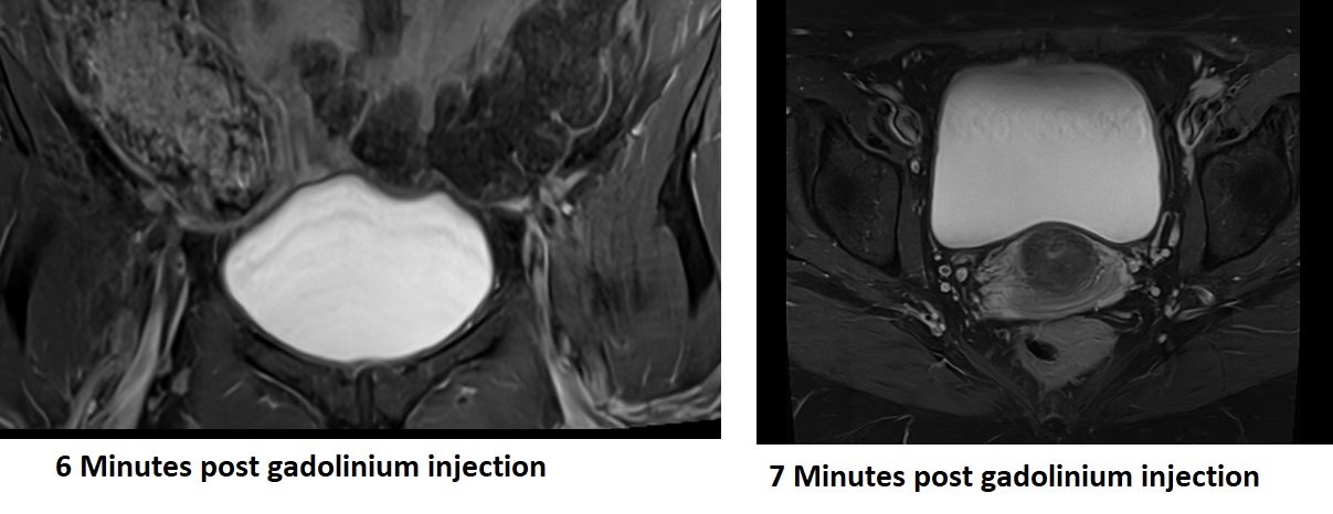
The Gadolinium Pseudo Layering artifact can occur in urinary bladder MRI when contrast-enhanced images are obtained. This artifact manifests as a pseudo-layering or stratified appearance of the gadolinium contrast agent within the urinary bladder.
The artifact is caused by the settling of the contrast agent in the dependent portion of the bladder due to gravity. As a result, the contrast agent accumulates more in certain areas, creating a layered appearance within the bladder lumen.
The Gadolinium Pseudo Layering artifact in urinary bladder MRI can be problematic as it can mimic the presence of actual layers or lesions within the bladder. This can lead to misinterpretation of the images and potential false-positive findings.


Here are some strategies to minimize or avoid Gadolinium Pseudo Layering Artifact
Dynamic imaging: Employing dynamic imaging techniques can be beneficial in visualizing the temporal changes in contrast agent distribution within the bladder. Serial acquisitions over time can provide a more accurate representation of the bladder anatomy and help differentiate between true pathology and the pseudo-layering artifact.

Timing of image acquisition: Timing the image acquisition appropriately is crucial to capture the contrast agent when it is well-mixed within the bladder. Acquiring images after allowing sufficient time for the contrast agent to diffuse and distribute evenly can help reduce the impact of the artifact.
References:
- Mai, W. (2008). Pseudolayering Artefact on Postcontrast Magnetic Resonance Images of the Bladder of 18 Dogs and Three Cats. Vet Rec, 163(4), 117-119. doi:10.1136/vr.163.4.116.
- Auffermann, W.F., et al. (2007). Gadolinium-DTPA-induced pseudolayering artifact on magnetic resonance imaging of the liver. Investigative Radiology, 22(5), 414-417.
- Murata, N., et al. (2016). Gadolinium retention after contrast-enhanced MRI. JAMA, 316(7), 700-702.