TI scout (look-locker) MRI
A TI-scout (TI refers to inversion time) is a type of imaging sequence used in cardiovascular magnetic resonance (CMR) to assess the nulling time of normal myocardium after the administration of a gadolinium-based contrast agent. This sequence is crucial for determining the optimal inversion time to null the signal from specific tissues, such as the myocardium, during the delyed PSIR image aqusition.
Here’s a breakdown of how it works:
- Inversion Time (TI): This refers to the delay time after which an MRI sequence is applied following the inversion of magnetization. In the TI scout sequence, multiple images are acquired with different TIs to allow the MRI to capture images at different points of recovery of the longitudinal magnetization.
- Purpose of the TI Scout: In cardiac MRI, particularly when using sequences like Delayed Enhancement or Late Gadolinium Enhancement (LGE), it is crucial to find the inversion time that causes the normal myocardium to appear nulled (dark) in the images. This enhances the contrast between healthy myocardial tissue and pathological tissues (like fibrosis or scars), which do not null at the same TI.
- Process: During a TI scout sequence, multiple images are acquired at different inversion times (ranging from 90 to 800 milliseconds, as in your example). This range is used to progressively see how the myocardium signal intensity changes as the inversion time increases.
- Optimal Nulling Point: By reviewing these images, the radiologist or technician can determine the specific inversion time at which the normal myocardium appears darkest (nulled), while pathological tissues retain higher signal intensity and appear brighter. This inversion time is then used for the late gadolinium enhancement (LGE) imaging sequences to ensure optimal contrast for visualization and assessment of myocardial tissue health.
Image Appearance
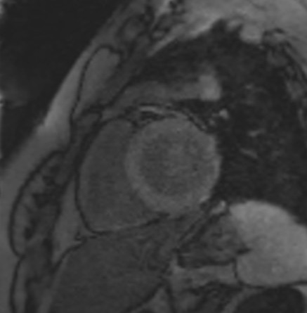
TI Range 90-120 milliseconds:
- Blood Appearance: At this short inversion time, blood typically appears dark. This is because the longitudinal magnetization of blood has not yet recovered sufficiently to provide a bright signal.
- Myocardium Appearance: The myocardium appears gray to bright in this range. The brightness indicates that the myocardium’s longitudinal magnetization is in the process of recovering more rapidly than that of blood, but has not reached full recovery.
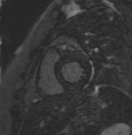
TI Range 200-280 milliseconds:
- Blood Appearance: Blood appears bright at this mid-range TI. This brightness occurs because the blood’s magnetization has had enough time to recover substantially, leading to a strong signal in the MRI images.
- Myocardium Appearance: The myocardium appears dark. This suggests that the TI is near or at the point of optimal nulling for healthy myocardial tissue, where its signal is minimized due to complete or nearly complete recovery of magnetization.
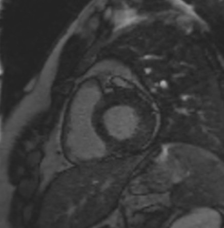
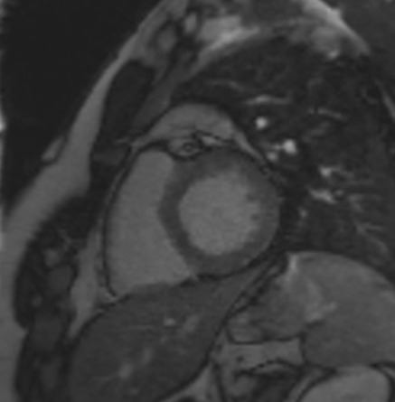
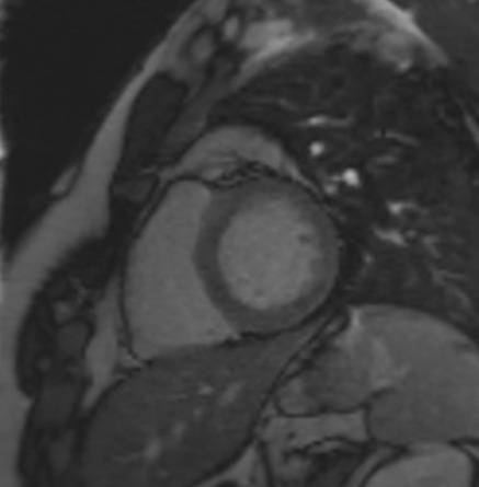
Long TI (400-800 milliseconds):
- Blood: Continues to appear bright as the inversion time increases. With longer TIs, blood has fully relaxed back to its baseline state, maximizing its signal and thus appearing bright in MRI images.
- Myocardium: Appears gray in this range. After the myocardium has passed its null point (typically around 200-280 milliseconds), it gradually begins to regain some signal as the magnetization continues to relax towards its baseline, resulting in a gray appearance.
TI 90
TI 120

TI 150

TI 200
TI 250

TI 280
TI 300

TI 350
TI 400
TI 450

TI 500

TI 600

References
- Ogawa, M., Matsumura, Y., & Tsuchihashi, T. (2011). Characteristics of TI scout image (look-locker) in the myocardial delayed enhancement MRI and the consistency of the null point between look-locker and IR-T1TFE method. Nihon Hoshasen Gijutsu Gakkai Zasshi, 67(6), 666-672. https://doi.org/10.6009/jjrt.67.666
- Bannan, B., Aguet, J., House, A. V., Gill, N., Tassos, V. P., Amirabadi, A., Seed, M., Lam, C. Z., & Yoo, S.-J. (2021). Usefulness of TI-scout images in the assessment of late gadolinium enhancement in children. Journal of Cardiovascular Magnetic Resonance, 23(28). https://doi.org/10.1186/s12968-021-00718-9
Pandey, T., Jambhekar, K., Shaikh, R., Lensing, S., & Viswamitra, S. (2013). Utility of the inversion scout sequence (TI scout) in diagnosing myocardial amyloid infiltration. International Journal of Cardiovascular Imaging, 29(1), 103-112. http://doi.org/10.1007/s10554-012-0042-4