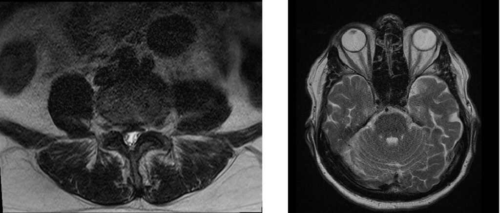
Signal-to-Noise Ratio(SNR) related Artifact
Signal-to-noise ratio (SNR)-related artifacts in MRI refer to the image distortions that result from low SNR levels in the acquired images. When the SNR is low, the images become noisy and may appear grainy or pixelated, making it difficult to distinguish small details. In addition to the reduction in image quality, low SNR can also lead to other artifacts, such as blurring, loss of contrast, and ghosting.
To avoid SNR-related artifacts in MRI, it is essential to optimize the acquisition parameters to obtain the highest possible SNR. This can be achieved by increasing the signal strength, reducing the noise level, and optimizing the receiver bandwidth.
Here are some strategies to minimize or avoid Signal-to-noise ratio-related artifact :
Use the appropriate coil: Using a suitable coil for the anatomy of interest can help increase the SNR.

Increase the number of averages: By acquiring multiple images and averaging them, the SNR can be improved.

Optimize the receiver bandwidth: Adjusting the receiver bandwidth to match the signal frequency can help to increase the SNR.

Increase the magnetic field strength: Higher magnetic field strengths provide increased signal-to-noise ratios.

Use noise-reducing techniques: Applying noise-reducing filters or denoising algorithms can help to improve the SNR.
Field of View (FOV): Choose an appropriate FOV that covers the region of interest without unnecessary inclusion of background noise. A smaller FOV concentrates the signal, leading to better SNR.

Matrix size : Optimize the matrix size based on the desired spatial detail and SNR requirements. A higher matrix size increases spatial resolution but can reduce SNR. Strike a balance between the two.

Echo time (TE) and repetition time (TR): Select appropriate TE and TR values for the specific imaging technique and desired contrast. Shorter TE values can provide better T1-weighted contrast, while longer TE values are suitable for T2-weighted contrast. Optimize TE and TR based on tissue properties and the desired contrast-to-noise ratio.
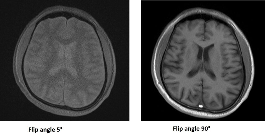
Flip angle: Choose the flip angle that maximizes the tissue signal while considering the specific imaging sequence and T1 relaxation times of the tissues. Be cautious of saturation effects at high flip
References:
- Madore, B. (2010). Techniques for improving SNR in MRI: a review. Magnetic Resonance in Medicine, 63(3), 701-714.
- Noll DC, Pauly JM, Meyer CH. (1992). “Multidimensional radiofrequency pulse design for magnetic resonance imaging”. Medical Physics, 19(6), 1261-127
- Bernstein, M.A., King, K.F., & Zhou, X.J. (2004). Handbook of MRI Pulse Sequences. Elsevier Health Sciences.