FLASH/SPGR/T1-FFE/RF Spoiled SARGE/RSSG/FAstFE/T1-FAST
FLASH MRI, known as Fast Low Angle Shot MRI, is a prevalent gradient-echo sequence in imaging, especially for abdominal studies. This rapid imaging technique employs low flip angle RF pulses, typically less than 90 degrees, paired with a reading gradient reversal to generate a gradient-echo signal. Designed explicitly for swift image capture, FLASH MRI is crucial in scenarios where movement might compromise image quality, such as dynamic studies. The reduced flip angle ensures a quick return to equilibrium of longitudinal magnetization, while a strong spoiler gradient eliminates transverse magnetization. This results in both T1-weighted and T2*-weighted contrasts, making FLASH MRI indispensable for various clinical applications, including angiography, cardiac imaging, and notably, abdominal imaging.
FLASH MRI Physics
The foundational principle of FLASH MRI is based on gradient echo sequences. The RF pulse in FLASH is tilted at a low flip angle instead of 90°, which means that the time required to return to equilibrium is reduced, allowing faster imaging. The echo is formed by gradient reversal rather than the application of a 180° RF pulse, which is the case in traditional spin-echo sequences. This gradient reversal causes dephased protons to realign. Since the recovery of longitudinal magnetization is rapid, due to the small flip angles, subsequent excitations can occur in quick succession, enabling faster image acquisition.
MRI Image Appearance of FLASH Image
Dynamic Imaging: Due to its rapid acquisition capabilities, FLASH MRI is ideal for dynamic studies, such as liver and breast imaging, as well as real-time joint motion capture.
Angiography: FLASH MRI’s ability to capture rapid sequences makes it useful for contrast-enhanced magnetic resonance angiography (MRA) of various body parts.
Abdominal Imaging: FLASH MRI offers faster imaging options, reducing motion artifacts caused by breathing or bowel movements.
Cardiac Imaging: FLASH’s speed is advantageous for imaging the constantly moving heart and capturing different phases of the cardiac cycle.
FLASH MRI Applications
Dynamic Imaging: Due to its rapid acquisition capabilities, FLASH MRI is ideal for dynamic studies, such as liver and breast imaging, as well as real-time joint motion capture.
Angiography: FLASH MRI’s ability to capture rapid sequences makes it useful for contrast-enhanced magnetic resonance angiography (MRA) of various body parts.
Abdominal Imaging: FLASH MRI offers faster imaging options, reducing motion artifacts caused by breathing or bowel movements.
Cardiac Imaging: FLASH’s speed is advantageous for imaging the constantly moving heart and capturing different phases of the cardiac cycle.
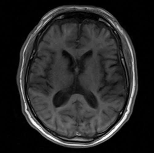
Tissues and their T1 FLASH appearance
Brain:
- CSF: Dark.
- Fat: Bright.
- White Matter: Intermediate to bright.
- Gray Matter: Intermediate to dark.
- Bone (skull): Dark.
- Bone Marrow: Intermediate to bright.
- Blood Vessels: Mostly Dark, Depending on flow characteristics, can be bright or dark
- Pituitary Gland: Intermediate.
- Choroid Plexus: Intermediat.
- Cerebellum: Intermediate.
- Brain Stem: Intermediate.
- Sinuses: Dark (air-filled).
- Thalamus, Putamen, Hippocampus, Caudate Nucleus: Intermediate.
- Corpus Callosum: Intermediate.
- Pineal Gland: Intermediate.
Spine:
- Spinal Cord: Intermediate.
- CSF: Dark.
- Bone: Dark.
- Bone Marrow: Intermediate to bright.
- Intervertebral Disc: Intermediate.
- Ligaments: Intermediate to Dark.
- Nerve Roots: Intermediate.
Abdomen and Pelvis:
- Liver: Intermediate.
- Gallbladder and Common Bile Duct: Dark.
- Spleen: Intermediate.
- Kidney: Intermediate.
- Ureters: Intermediate.
- Pancreas: Intermediate.
- Urinary Bladder: Bright.
- Prostate: Intermediate.
- Uterus: Intermediate to bright.
Musculoskeletal:
- Muscle: Intermediate to dark.
- Bone: Dark (low signal).
- Bone Marrow: Intermediate to bright.
- Blood Vessels: Mostly Dark.
- Fat: bright.
- Ligaments: Intermediate to dark.
- Nerve Roots: Intermediate.
- Cartilage: Intermediate to dark
- Synovial Fluid: Dark.
- Tendons: Intermediate to dark.
Use
- Very useful for breast imaging
- Useful for abdominal imaging
- Useful for small bowel and MR colonoscopy imaging
- Useful for chest imaging
- Useful for brain imaging (to acquire fast scans in uncooperative patients)
- Useful for adrenal imaging
- Useful for angiography
- Useful for liver imaging
*Most of the abdominal imaging uses fat saturated FLASH sequences *
flash axial sequence used in uncooperative patients brain imaging
flash coronal sequence used in subclavian angiography imaging

FLASH coronal sequence used in abdominal angiography imaging

Flash coronal sequence used in upper legs angiography imaging

flash coronal sequence used in lower legs angiography imaging
