True FISP/ FIESTA / Balanced SARGE / BASG / True SSFP / STERF
The T2 True Fast Imaging with Steady State Precession (TrueFISP) or Fast Imaging Employing Steady-state Acquisition (FIESTA) MRI sequence is an advanced imaging technique that capitalizes on a fully balanced gradient waveform. One of its distinguishing features is that the image contrast it produces is based on T1/T2 ratios. Nonetheless, when both the repetition time (TR) and echo time (TE) are kept short, the T1 contrast component tends to stay constant. This results in images that are predominantly T2-weighted.
A notable characteristic of TrueFISP is its heightened sensitivity to magnetic field inhomogeneities. This can sometimes manifest as interference stripes or Moiré fringes in the resultant images. To reduce this susceptibility, it’s recommended to maintain the TR very short and to execute a shimming procedure, which is a method to correct field inhomogeneities, prior to the image acquisition.
One of the significant advantages of this sequence is its rapid acquisition speed. This quick capture rate, combined with its minimal sensitivity to patient motion, makes TrueFISP especially reliable. Such attributes are beneficial in clinical scenarios, especially when imaging patients who might find it challenging to hold their breath or remain still during the scan.
MRI image appearance of True FISP/ FIESTA
The easiest way to identify TrueFISP images is by looking for blood vessels and fluid-filled spaces in the body. Examples include cerebrospinal fluid in the brain’s ventricles and spinal canal, free fluid in the abdomen, fluid in the gall bladder and common bile duct, synovial fluid in joints, fluid in the urinary tract and bladder, edema, and any other pathological fluid collections in the body. In TrueFISP images, fluids and blood vessels typically appear bright.
Tissues and their TrueFISP appearance
Brain:
- CSF: Bright.
- Fat: Bright.
- White Matter: Dark.
- Gray Matter: Intermediate to Dark.
- Bone (skull): Dark.
- Bone Marrow: Intermediate to bright.
- Blood Vessels: Mostly bright.
- Pituitary Gland: Intermediate.
- Choroid Plexus: Intermediate.
- Cerebellum: Intermediate to dark.
- Brain Stem: Intermediate to dark.
- Sinuses: Dark (air-filled).
- Thalamus, Putamen, Hippocampus, Caudate Nucleus: Intermediate.
- Corpus Callosum: Dark.
- Pineal Gland: Intermediate.
Spine:
- Spinal Cord: Intermediate.
- CSF: Bright.
- Bone: Dark.
- Bone Marrow: Intermediate to bright.
- Intervertebral Disc: Intermediate
- Ligaments: Intermediate to Dark.
- Nerve Roots: Intermediate.
Abdomen and Pelvis:
- Liver: Intermediate to dark.
- Gallbladder and Common Bile Duct: Bright
- Spleen: Intermediate to dark.
- Kidney: Intermediate.
- Ureters: Intermediate.
- Pancreas: Intermediate to dark.
- Urinary Bladder: Bright.
- Prostate: Intermediate.
- Uterus: Intermediate.
Musculoskeletal:
- Muscle: Intermediate to dark
- Bone: Dark (low signal)
- Bone Marrow: Intermediate to bright
- Blood Vessels: Mostly bright.
- Fat: Dark
- Ligaments: Intermediate to dark
- Nerve Roots: Intermediate
- Cartilage: Intermediate to dark
- Synovial Fluid: Bright
- Tendons: Intermediate to dark
Use
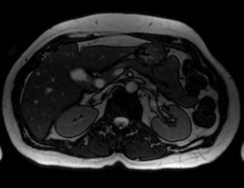
- Very useful for abdominal imaging
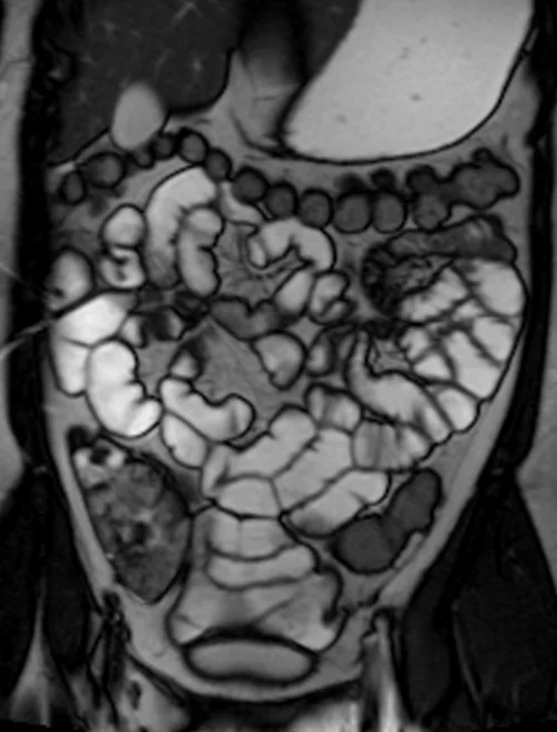
- Very useful for small bowel and MR colonoscopy imaging
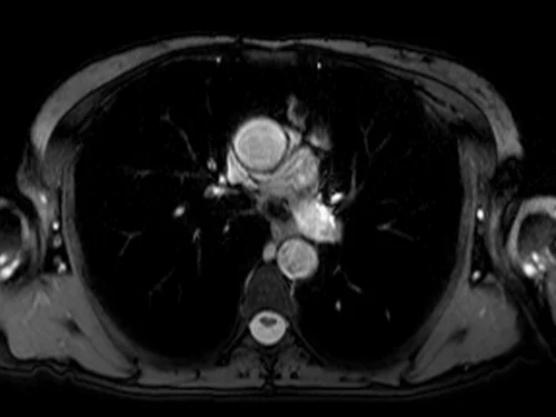
- Very useful for chest imaging
- Useful for brain imaging (to acquire fast scans in uncooperative patients)
- Useful for adrenal imaging
- Useful for pancreas imaging
- Useful for liver imaging
True FISP axial sequence used in uncooperative patients' brain imaging

True FISP axial sequence used in chest imaging
True FISP axial sequence used in abdomen imaging
True FISP coronal sequence used in enterography mri imaging
True FISP coronal sequence used pancreas imaging
