MRI Defecography (Proctogram)
Indications for mri Defecography (Proctogram) scan
- Descending perineum syndrome
- Internal rectal intussusception
- Difficulty in opening bowel
- Obstructed defecation
- Faecal incontinence
- Pelvic floor dysfunction
- Rectal prolapse
- Sigmoidocele
- Rectocele
- Enterocele
- Anismus
Contraindications
- Any electrically, magnetically or mechanically activated implant (e.g. cardiac pacemaker, insulin pump biostimulator, neurostimulator, cochlear implant, and hearing aids)
- Intracranial aneurysm clips (unless made of titanium)
- Pregnancy (risk vs benefit ratio to be assessed)
- Ferromagnetic surgical clips or staples
- Metallic foreign body in the eye
- Metal shrapnel or bullet
Patient preparation for Defecography (Proctogram)
- It is important that the bowel is clean and empty before the procedure. Patient should be supplied with a laxative suppository and instructions leaflet for how to insert the suppository.
- The suppository should insert into the rectum on the morning of the test. Instruct the patient to retain the suppository for 15 minutes and then empty your bowel completely.
- A satisfactory written consent form must be taken from the patient before entering the scanner room
- Ask the patient to remove all metal object including keys, coins, wallet, any cards with magnetic strips, jewellery, hearing aid and hairpins
- Ask the patient to undress and change into a hospital gown
- If possible provide a chaperone for claustrophobic patients (e.g. relative or staff )
- Offer earplugs or headphones, possibly with music for extra comfort
- Explain the procedure to the patient
- Instruct the patient to keep still
- Note the weight of the patient
Positioning for Defecography (Proctogram)
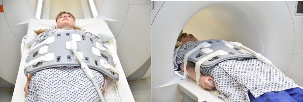
- Before positioning the patient place some inco pads over the table (over the spine coil) and cover it with a disposable couch roll. Now position the patient over the couch roll in supine position with head pointing towards the magnet (head first supine)
- Position the patient over the spine coil and place the body coil over the pelvis(two inches above iliac crest down to three inches below symphysis pubis)
- Securely tighten the body coil using straps to prevent respiratory artefacts
- Give a pillow under the head for extra comfort (do not give cushions under the legs )
- Centre the laser beam localiser over anterior superior iliac spine (2 inches below iliac crest)
Recommended MRI Defecography (Proctogram) Protocols and Planning
MRI Defecography localiser
A three-plane localizer must be taken at the beginning to localize and plan the sequences. Localizers are normally less than 25 seconds and are T1\T2-weighted low-resolution scans.
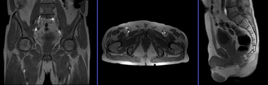
T2 tse sagittal 3mm SFOV pre proctogram
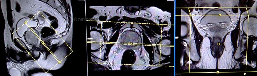
Plan the sagittal slices on the axial plane; angle the positioning block parallel to the interpubic fibrocartilage and the anal canal. Check the positioning block in the other two planes. An appropriate angle must be given in the coronal plane (parallel to the rectum and the anal canal). Slices must be sufficient to cover the whole pelvis from the right acetabulum to the left acetabulum. FOV must be big enough to cover the whole pelvis (normally 270mm-300mm). Adding saturation bands on top and front of the sagittal block will reduce artifacts from arterial pulsation and breathing.
Parameters
TR 4000-5000 | TE 100-120 | SLICE 3 MM | FLIP 130-150 | PHASE A>P | MATRIX 320X320 | FOV 270-300 | GAP 10% | NEX(AVRAGE) 3 |
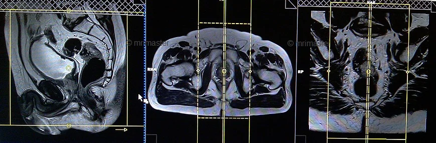
T2 tse axial oblique 3mm SFOV pre proctogram
Plan the axial oblique slices on the sagittal plane; angle the positioning block perpendicular to the anal canal. Check the positioning block in the other two planes. An appropriate angle must be given in the coronal plane (perpendicular to the anal canal). Slices must be sufficient to cover the entire anal canal.
Parameters
TR 4000-5000 | TE 100-120 | SLICE 3 MM | FLIP 130-150 | PHASE A>P | MATRIX 320X320 | FOV 180-230 | GAP 10% | NEX(AVRAGE) 4 |
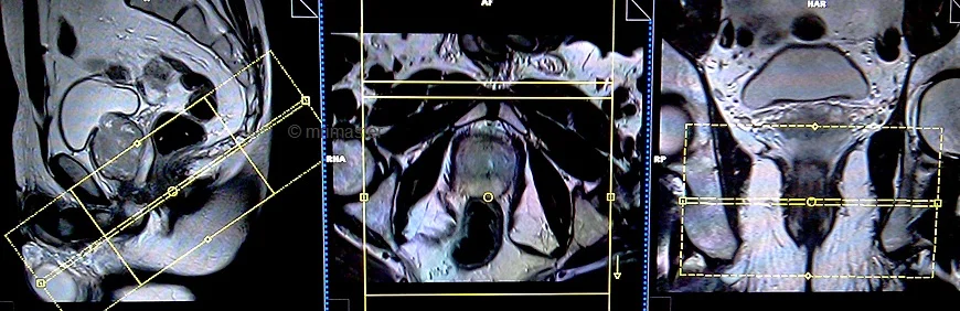
T2 tse coronal oblique 3mm SFOV pre proctogram
Plan the coronal oblique slices on the sagittal plane; angle the positioning block parallel to the anal canal. Check the positioning block in the other two planes. An appropriate angle must be given in the axial plane (horizontally across the anal canal or parallel to the right and left hip joint). Slices must be sufficient to cover the whole anal canal.
Parameters
TR 3000-4000 | TE 100-120 | SLICE 3 MM | FLIP 130-150 | PHASE R>L | MATRIX 320X256 | FOV 180-230 | GAP 10% | NEX(AVRAGE) 5 |
pause for proctogram
Next, guide the patient to lie down in either a right or left lateral position. The radiologist will gently insert a small catheter lubricated with ky jelly into the rectum and inject a small amount of sonography gel. Afterward, remove the catheter and place the patient back into the initial position. Instruct the patient to clench, relax, and strain during image capture.
localiser
A three-plane localizer must be taken at the beginning to localize and plan the sequences. Localizers are normally less than 25 seconds and are T1\T2-weighted low-resolution scans.
T2 TRUFISP cine, small FOV 10mm, 1 second, 150 measurements.
Plan the sagittal slices on the axial plane; angle the positioning block parallel to the interpubic fibrocartilage and the anal canal. Check the positioning block in the other two planes. An appropriate angle must be given in the coronal plane (parallel to the rectum and the anal canal). Slice must be positioned over the middle of the rectum and anal canal.
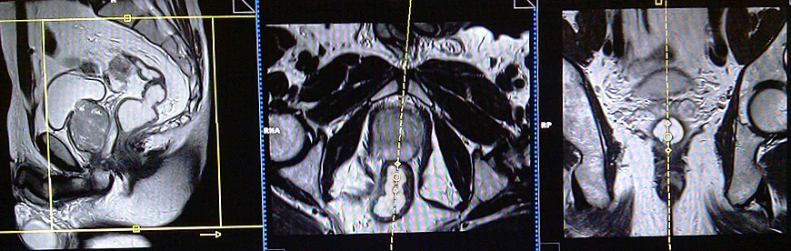
After approximately five measurements, instruct the patient to strain and expel the gel from the rectum onto the incopads beneath, while ensuring that cine scans are continuously running during this process.
Parameters
TR 40-50 | TE 1-2 | SLICE 5-10 MM | FLIP 70 | PHASE A>P | MATRIX 208X208 | FOV 250-300 | OVERS. 50% | CINE 150measurement |