MRI Partial Fourier
Partial Fourier
Partial Fourier in MRI (Magnetic Resonance Imaging) refers to a data acquisition strategy used to reduce the time required for acquiring a complete MRI image. The MRI image acquisition process involves gathering multiple sets of data points, known as “k-space” data, which are subsequently transformed into an image using a mathematical technique called Fourier transformation.
In conventional MRI data acquisition, the entire k-space is populated by acquiring all the necessary data points. However, acquiring all these data points within k-space can be time-consuming, especially for high-resolution or 3D imaging, resulting in longer scan durations. Partial Fourier is a method employed to expedite the acquisition process while still producing satisfactory images.
Within Partial Fourier imaging, only a segment of the k-space is procured, typically along one dimension. The absent data in the remaining dimension(s) are approximated or filled using mathematical algorithms. The Partial Fourier technique capitalizes on the symmetrical attributes of both k-space and image space to accurately predict the missing data. This facilitates quicker acquisition times, thereby diminishing the overall scan duration.
Partial Fourier imaging proves particularly beneficial in scenarios necessitating reduced scan times, such as when imaging mobile structures (e.g., the heart and small bowel), during breath-hold examinations to curtail scan duration, or when patients experience difficulty maintaining stillness over extended periods.
Partial Fourier off

Scan time 2.30min
Partial Fourier 6/8

Scan time 1.40min
Partial Fourier 4/8

Scan time 1.00min
Advantages of Partial Fourier
Faster Scan Times: The primary advantage of Partial Fourier imaging is the reduction in scan time. By acquiring only a portion of the k-space data, the overall scan duration is shortened, making the MRI examination more tolerable for patients. This is particularly important for patients who may have difficulty holding their breath or staying still for extended periods.
Motion Artifact Reduction: Since Partial Fourier imaging is particularly useful for dynamic studies or when imaging moving structures, it helps minimize motion artifacts. By acquiring data faster, there’s less chance for motion-related distortions to affect the image quality.
Flexible Breath-Hold Imaging: For breath-hold exams, such as abdominal or cardiac imaging, Partial Fourier allows for the collection of sufficient data in a shorter breath-hold time, which is crucial for patient cooperation and image quality.
Disadvantages Partial Fourier
Reduced Signal-to-Noise Ratio (SNR): The reduction in acquired data can result in a lower SNR, affecting image quality. In areas where the missing data is estimated, the accuracy might be compromised, leading to decreased SNR and lower image contrast.
Image Artifacts: One of the main disadvantages of Partial Fourier imaging is the potential for artifacts. The mathematical assumptions required for accurate estimation of the missing k-space data may not always hold true, leading to artifacts that degrade image quality.
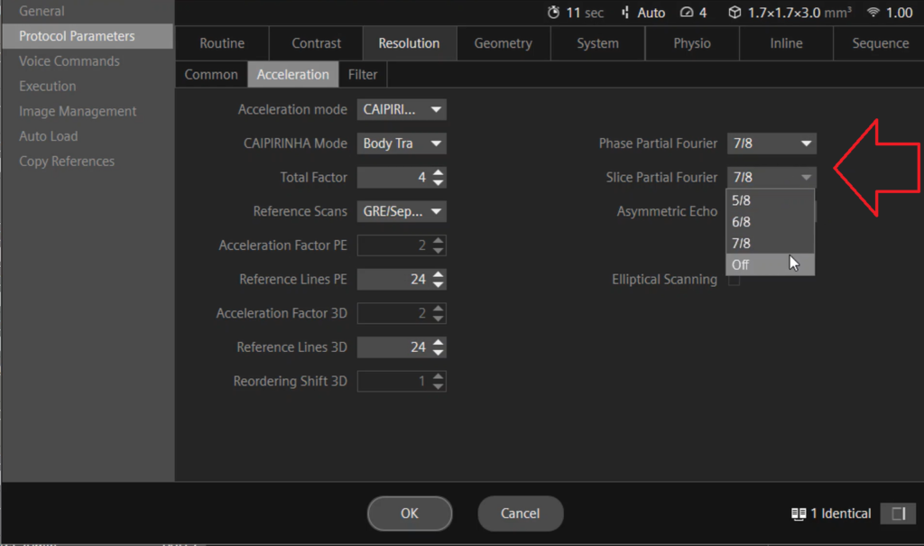
Where can partial Fourier be found and how is it used?
Within a Siemens MRI scanner, the “resolution” tab under the “acceleration” category hosts the Partial Fourier setting, allowing you to toggle it between on and off. Activating this option presents various Partial Fourier choices, such as 7/8, 6/8, or 5/8. Opting for 7/8, for instance, results in 87% of the K space being filled with acquired data, while mathematical algorithms fill the remaining portion. In contrast, selecting 5/8 fills only 62% of the K space with acquired data, with mathematical algorithms completing the remainder. The appropriate selection should align with the specific clinical requirements.
References
- Lustig, M., Pauly, J. M., & Pipe, J. G. (2002). A method for undersampled 3DFT MR angiography. Magnetic Resonance in Medicine, 49(6), 1219-1227. doi:10.1002/mrm.10165
- Pipe, J. G., Zwart, N. R., Aboussouan, E. A., & Robison, R. K. (2002). Partial Fourier reconstruction in MRI. Medical Physics, 29(4), 755-764. DOI: 10.1118/1.1468977
- Trzasko, J. D., & Manduca, A. (2001). A geometric interpretation of partial Fourier imaging. Magnetic Resonance in Medicine, 46(4), 789-798. DOI: 10.1002/mrm.1247
- Kellman, P., Epstein, F. H., McVeigh, E. R., & Lima, J. A. (2001). Reduction of motion and flow artifacts in triggered MRI using partial-Fourier reconstruction. Magnetic Resonance in Medicine, 45(2), 289-297. DOI: 10.1002/1522-2594(200102)45:2<289::AID-MRM1020>3.0.CO;2-L