MRI Fingers : Protocol and Planning
Indications for fingers MRI scan
- Marrow abnormalities (e.g. bone contusions, osteonecrosis, marrow oedema syndromes, and stress fractures)
- Synovial based disorders ( e.g. synovitis, tenosynovitis, bursitis, and ganglion cysts)
- Infections of bone, joint, or soft tissue (eg. osteomyelitis, osteo arthritis )
- Neoplasms of bone, joint or soft tissue
- Avascular necrosis
- Nerve Impingement
- Fractures in children
- Soft-tissue masses
- Occult fracture
- Ganglion cyst
- Ligament tear
Contraindications
- Any electrically, magnetically or mechanically activated implant (e.g. cardiac pacemaker, insulin pump biostimulator, neurostimulator, cochlear implant, and hearing aids)
- Intracranial aneurysm clips (unless made of titanium)
- Pregnancy (risk vs benefit ratio to be assessed)
- Ferromagnetic surgical clips or staples
- Metallic foreign body in the eye
- Metal shrapnel or bullet
Patient preparation for MRI Fingers
- A satisfactory written consent form must be taken from the patient before entering the scanner room
- Ask the patient to remove all metal objects including keys, coins, wallet, cards with magnetic strips, jewellery, hearing aid and hairpins
- If possible provide a chaperone for claustrophobic patients (e.g. relative or staff )
- Offer earplugs or headphones, possibly with music for extra comfort
- Explain the procedure to the patient
- Instruct the patient to keep still
- Note the weight of the patient
Positioning for MRI Fingers
- Head first prone with arm up (superman position)
- Position the hand in the and and wrist coil or the large flex coil and immobilize it with cushions.
- Give cushions under the chest for extra comfort
- Centre the laser beam localiser over the metacarpophalangeal joint
- Register the patient o the scanner as 'head first supine'

Recommended MRI Fingers Protocols and Planning
Localiser
A three-plane localizer must be taken at the beginning to localize and plan the sequences. Typically, these localizers take less than 25 seconds and can be achieved using T1 weighted low-resolution scans. It is advisable to obtain additional localizers until you have acquired accurate axial, coronal, and sagittal localizer images.

T2 stir axial 3mm SFOV
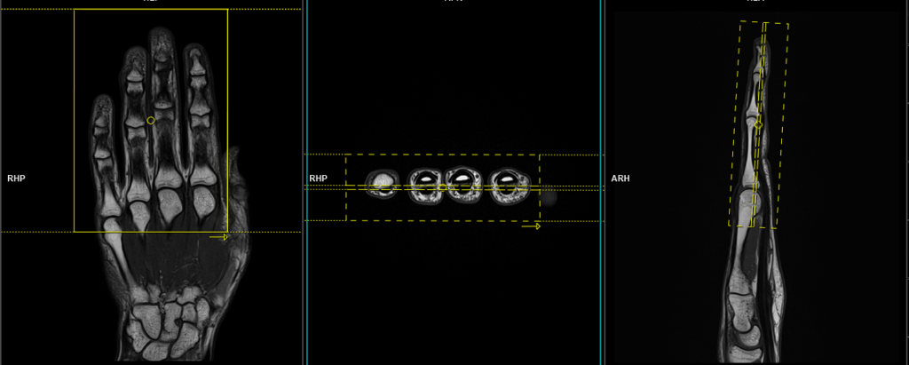
Plan the axial slices on the coronal localizer and angle the positioning block perpendicular to the phalangeal bones. Check the positioning block in the other two planes. An appropriate angle must be used in the sagittal plane, perpendicular to the phalangeal bones. The slices should be sufficient to cover the entire hand, from the tip of the fingers to the line of the distal radioulnar joint.

Parameters
TR 5000-6000 | TE 110 | FLIP 150 | NEX 2 | SLICE 3MM | MATRIX 288X288 | FOV 100-130 | PHASE A>P | GAP 10% | TI 160 |
T1 tse axial 3mm SFOV
Plan the axial slices on the coronal localizer and angle the positioning block perpendicular to the phalangeal bones. Check the positioning block in the other two planes. An appropriate angle must be used in the sagittal plane, perpendicular to the phalangeal bones. The slices should be sufficient to cover the entire hand, from the tip of the fingers to the line of the distal radioulnar joint.
Parameters
TR 400-600 | TE 15-25 | SLICE 3 MM | FLIP 150 | PHASE A>P | MATRIX 320X320 | FOV 100-130 | GAP 10% | NEX(AVRAGE) 2 |
T1 tse coronal 2mm SFOV
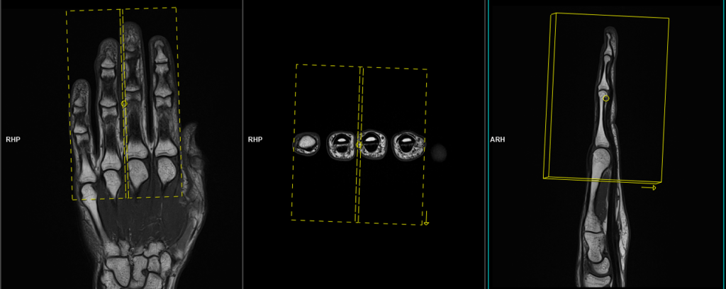
Plan the coronal slices on the axial plane, angling the positioning block parallel to the line across the phalangeal bones. Check the positioning block in the other two planes. An appropriate angle must be used in the sagittal plane, parallel to the phalangeal bones. The slices must be sufficient to cover the entire hand from the dorsal to palmar aspect.
Parameters
TR 400-600 | TE 15-25 | SLICE 2 MM | FLIP 130 | PHASE R>L | MATRIX 320X320 | FOV 140-160 | GAP 10% | NEX(AVRAGE) 2 |
T2 stir coronal 2mm SFOV
Plan the coronal slices on the axial plane, angling the positioning block parallel to the line across the phalangeal bones. Check the positioning block in the other two planes. An appropriate angle must be used in the sagittal plane, parallel to the phalangeal bones. The slices must be sufficient to cover the entire hand from the dorsal to palmar aspect.
Parameters
TR 3000-4000 | TE 110 | FLIP 130 | NEX 2 | SLICE 2 MM | MATRIX 320X256 | FOV 140-160 | PHASE R>L | GAP 10% | TI 160 |
T2 stir sagittal 2mm SFOV
Plan the sagittal slices on the axial plane; angle the position block perpendicular to the line across the phalangeal bones. Check the positioning block in the other two planes. An appropriate angle must be used in the coronal plane (parallel to the phalangeal bones). Slices must be sufficient to cover the fingers from index finger to little finger.
Parameters
TR 4000-5000 | TE 110 | FLIP 150 | NEX 2 | SLICE 3MM | MATRIX 256X256 | FOV 140-160 | PHASE A>P | GAP 10% | TI 160 |