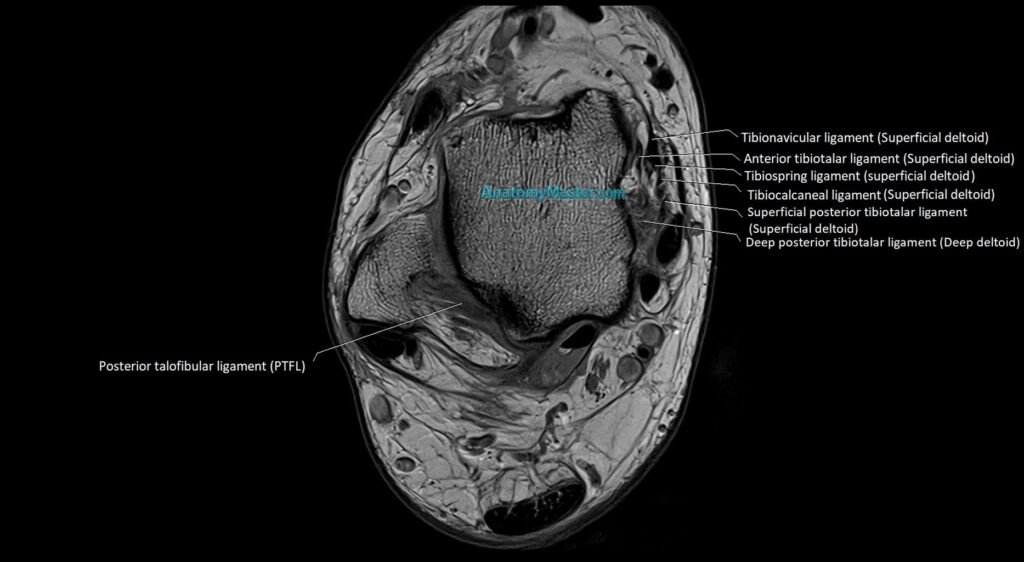
3T MRI Ankle Anatomy : High-resolution 1.5mm Deep Resolve Slices (Subscription-Based Premium Access)
Explore our advanced MRI ankle anatomy library, featuring ultra-high-resolution anatomical images of the ankle in axial, sagittal, and coronal planes. This specialist learning resource has been developed from carefully repeated volunteer scans using advanced 3T MRI scanners, dedicated high-channel coils, and AI-powered Deep Resolve image enhancement. The result is a clinically realistic MRI anatomy platform that demonstrates true anatomical detail, including the distal tibia, distal fibula, talus, calcaneus, ankle joint, subtalar joint, tendons, ligaments, nerves, vessels, cartilage, joint spaces, and surrounding soft tissues, while preserving the authenticity of real clinical MRI imaging.
The MRI ankle anatomy section includes high-resolution TSE T1-weighted images, carefully selected for anatomical clarity and educational value. Every individual slice has been manually labelled in a textbook-style format by our team, without the use of AI auto-labelling tools. This ensures high accuracy, consistent anatomical detail, and reliable learning support for radiologists, orthopaedic surgeons, foot and ankle surgeons, sports medicine clinicians, podiatrists, physiotherapists, radiology students, reporting radiographers, and anyone interested in detailed ankle anatomy.
Each labelled MRI image has been built into a smooth custom interactive scroller for easy learning. The interface is designed to be user-friendly on both desktop and mobile platforms. Double-clicking the scroller opens the image viewer in full-screen mode. The bottom toolbar allows users to switch between different imaging planes and use voice or text search to access detailed descriptions, including MRI and CT appearances of individual anatomical structures.
The side toolbar includes a powerful zoom function, allowing images to be magnified up to 10x without compromising image quality. This makes it easier to study fine anatomical structures and understand their relationship to surrounding ankle anatomy, including the Achilles tendon, tibialis posterior tendon, flexor hallucis longus tendon, peroneal tendons, anterior extensor tendons, tarsal tunnel, tibial nerve, posterior tibial vessels, lateral ligament complex, deltoid ligament, syndesmotic ligaments, and subtalar joint.
The labelling and anatomical content have been developed using multiple anatomy, orthopaedic, foot and ankle surgery, sports medicine, podiatry, musculoskeletal imaging, and radiology reference books to provide a highly detailed and clinically focused learning experience. The project has been created over five years by a dedicated team and is being authorised and verified by experienced radiologists to ensure accuracy and reliability.
The ankle MRI anatomy module is also supported by Level 1 and Level 2 dynamic quizzes. These quizzes generate new questions in every session, helping users actively test their knowledge and build long-term understanding of ankle anatomy.
This module is part of a separate radiology-focused anatomy website and is not available within the MRIMaster app. The full platform will be offered through an affordable annual subscription, with a strong focus on supporting students and healthcare professionals worldwide.
MRI Ankle axial 1.5mm Deep Resolve Labelled Image(sample)
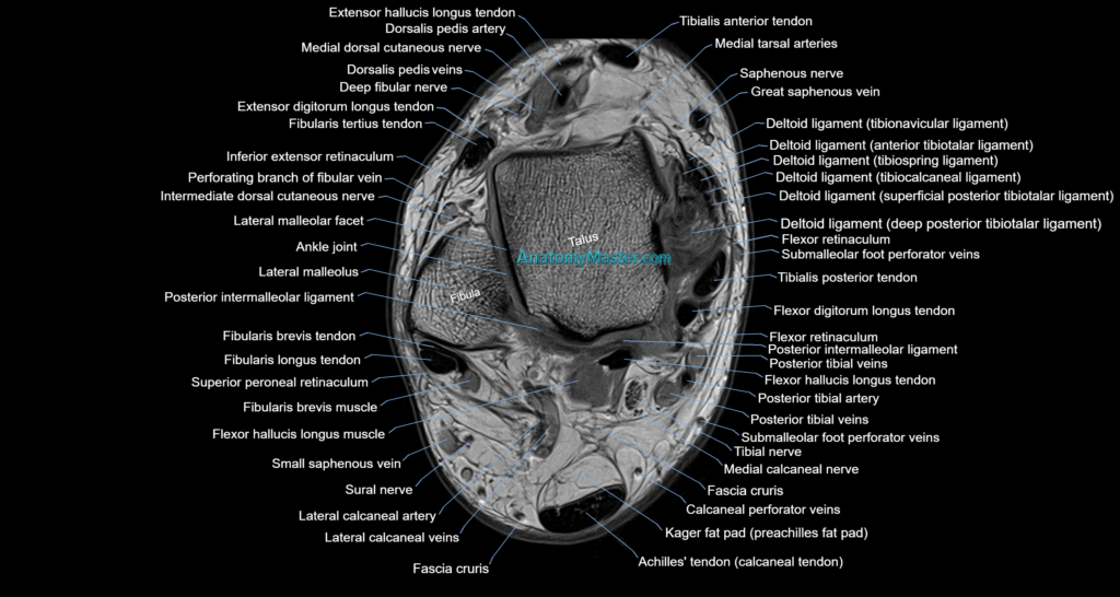
MRI Ankle sagittal 1.5mm Deep Resolve Labelled Image(sample)
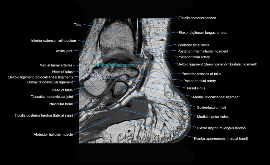
MRI Ankle coronal 1.5mm Deep Resolve Labelled Image(sample)
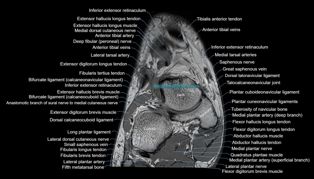
MRI Ankle ligaments axial 1.5mm Deep Resolve Labelled Image(sample)