Pituitary (Sella Turcica) MRI Protocols and Planning
Common Indications for MRI of the Pituitary
- Pituitary hypofunction, hormone hypersecretion,
- Visual field defects, bitemporal hemianopia
- Amenorrhea, galactorrhea, infertility
- Diplopia, ptosis, altered facial sensation
- Microadenomas and macroadenomas
- Delayed or frank absence of puberty
- Abnormal height and growth rate
- Decreased libido, impotence
- Nonspecific headache
- Tumour recurrence
- Hypoglycemia
- Dyspareunia
- Obesity
Contraindications
- Any electrically, magnetically or mechanically activated implant (e.g. cardiac pacemaker, insulin pump biostimulator, neurostimulator, cochlear implant, and hearing aids)
- Intracranial aneurysm clips (unless made of titanium)
- Pregnancy (risk vs benefit ratio to be assessed)
- Ferromagnetic surgical clips or staples
- Metallic foreign body in the eye
- Metal shrapnel or bullet
Please check our new video tutorial for protocols and planning
Patient preparation for pituitary mri
- A satisfactory written consent form must be taken from the patient before entering the scanner room
- Ask the patient to remove all metal objects including keys, coins, wallet, cards with magnetic strips, jewellery, hearing aid and hairpins
- If possible provide a chaperone for claustrophobic patients (e.g. relative or staff )
- Contrast injection risk and benefits must be explained to the patient before the scan
- Gadolinium should only be given to the patient if GFR is > 30
- Offer earplugs or headphones, possibly with music for extra comfort
- Explain the procedure to the patient
- Instruct the patient to keep still
- Note the hight and weight of the patient
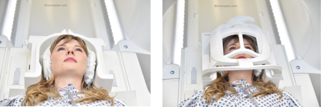
Positioning for Pituitary MRI
- Head first supine
- Position the head in the head coil and immobilise with cushions
- Give cushions under the legs for extra comfort
- Centre the laser beam localizer over the glabella
Recommended MRI Pituitary Protocols, Parameters, and Planning
Pituitary MRI localiser
A three-plane localizer must be taken at the beginning to localize and plan the sequences. Localizers are usually less than 25 seconds and are T1-weighted low-resolution scans.

T2 tse axial 4mm
Plan the axial slices on the sagittal plane and position the block parallel to the genu and splenium of the corpus callosum. Verify the planning block in the other two planes and ensure that an appropriate angle is maintained in the coronal plane, making it perpendicular to the line of the midline of the brain and the 4th ventricle. Ensure that the number of slices is sufficient to cover the entire brain from the vertex to the line of the foramen magnum.

Parameters
TR 4000-5000 | TE 100-120 | SLICE 4MM | FLIP 130-150 | PHASE R>L | MATRIX 320X320 | FOV 210-230 | GAP 10% | NEX(AVRAGE) 2 |
T1 tse DIXON 2mm sagittal SFOV
Plan the sagittal slices on the axial plane, and angle the planning block parallel to the midline of the brain. Verify the planning block in the other two planes. Ensure an appropriate angle is given in the coronal plane, parallel to the midline of the brain. Slices should be sufficient to cover the entire pituitary gland from the right to left internal carotid arteries.
Parameters
TR 400-500 | TE 15 | FAT SAT DIXON | NEX 4 | SLICE 2MM | MATRIX 256×256 | FOV 100-120 | PHASE A>P | OVERSAMPLE 100% | GAP 10% |
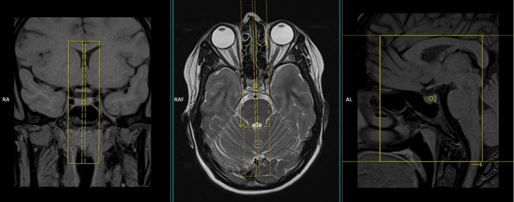
T2 tse coronal 2mm
Plan the coronal slices on the sagittal plane. Angle the planning block perpendicular to the Sella turcica. Check the planning block in the other two planes. An appropriate angle must be given in the axial plane (perpendicular to the midline of the brain). Slices must be sufficient to cover the whole pituitary gland from the anterior border of the sphenoid sinus to the line of the anterior border of the pons.

Parameters
TR 4000-5000 | TE 110 | FLIP 150 | NEX 4 | SLICE 2MM | MATRIX 256×256 | FOV 100-120 | PHASE R>L | OVERSAMPLE 100% | GAP 10% |
T1 tse DIXON 2mm coronal
Plan the coronal slices on the sagittal plane. Angle the planning block perpendicular to the Sella turcica. Check the planning block in the other two planes. An appropriate angle must be given in the axial plane (perpendicular to the midline of the brain). Slices must be sufficient to cover the whole pituitary gland from the anterior border of the sphenoid sinus to the line of the anterior border of the pons.

Parameters
TR 400-500 | TE 15 | FAT SAT DIXON | NEX 4 | SLICE 2MM | MATRIX 256×256 | FOV 100-120 | PHASE A>P | OVERSAMPLE 100% | GAP 10% |