High-Resolution CT Hip Anatomy: 1 mm Axial Images | Premium Subscription Access
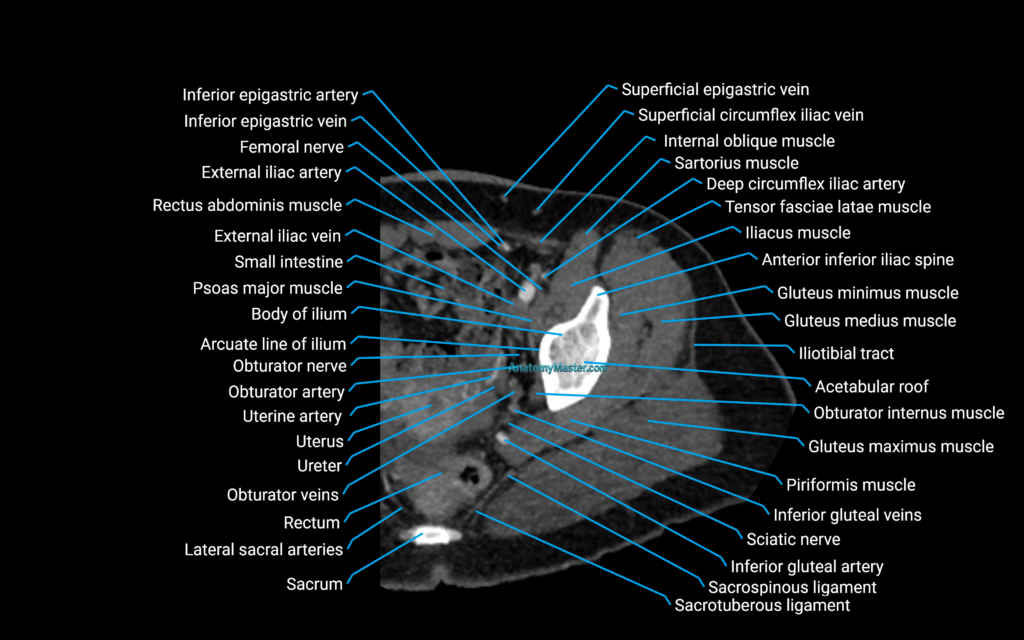
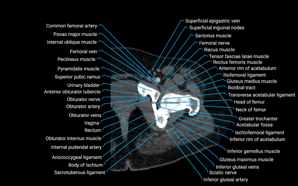
Explore our advanced CT hip anatomy library, featuring high-resolution anatomical images presented in the axial plane. This specialist learning module has been developed using thin-slice CT imaging to demonstrate clinically realistic cross-sectional anatomy of the hip joints and surrounding structures.
The CT hip anatomy section covers the bones, joints, muscles, tendons, ligaments, blood vessels, nerves and soft tissues of both hip regions. Important structures include the femoral head, femoral neck, greater and lesser trochanters, acetabulum, anterior and posterior acetabular walls, acetabular columns, pubis, ischium and ilium.
The module also demonstrates the hip joint space, joint capsule, acetabular labrum region, ligamentum teres, transverse acetabular ligament and surrounding fat planes. These anatomical relationships are particularly important when reviewing trauma, fractures, joint degeneration, postoperative imaging and preoperative planning.
The muscular anatomy includes the gluteus maximus, gluteus medius, gluteus minimus, iliopsoas, rectus femoris, sartorius, tensor fasciae latae, adductor muscles, obturator muscles, quadratus femoris and proximal hamstring muscles. Major tendons and their relationships to the hip joint and proximal femur are also demonstrated.
Important neurovascular structures include the femoral artery and vein, profunda femoris vessels, obturator vessels, sciatic nerve, femoral nerve and other nearby anatomical landmarks visible on CT.
Each axial CT image is manually labelled slice by slice in a textbook-style format, without the use of AI auto-labelling tools. This helps maintain anatomical accuracy and provides a reliable learning reference for radiologists, orthopaedic surgeons, trauma specialists, radiology trainees, reporting radiographers and students studying musculoskeletal and cross-sectional anatomy.
The labelled images are displayed within a smooth interactive scroller, allowing users to move naturally through the anatomy in the same way they would review a clinical axial CT examination. The platform works on desktop and mobile devices, while the full-screen viewer supports detailed examination of small anatomical structures.
The toolbar allows users to search for specific structures using voice or text search. This helps users quickly locate important landmarks such as the femoral head, acetabulum, femoral neck, greater trochanter, iliopsoas muscle, gluteal muscles, sciatic nerve and femoral vessels.
The side zoom tool provides up to 10× magnification without loss of image quality, allowing users to examine fine anatomical details and better understand the relationships between bones, joints, muscles, tendons, nerves, vessels and surrounding soft tissues.
This module is supported by dynamic Level 1 and Level 2 quizzes that generate new questions during every session. These interactive assessments help users actively revise, test their knowledge and retain complex CT hip anatomy.
CT Hip axial Anatomy: 1 mm Labelled Image (Sample)