CT Coronary Angiography Anatomy: High-Resolution Axial, 2-Chamber, 4-Chamber and Short-Axis Images | Premium Subscription Access
Explore our advanced CT coronary angiography anatomy library, featuring high-resolution, contrast-enhanced cardiac CT images displayed in axial, two-chamber, four-chamber and short-axis planes. This specialist learning module has been developed using clinically realistic coronary CT angiography images and high-quality multiplanar reconstructions to demonstrate the detailed anatomy of the heart, coronary arteries and surrounding cardiovascular structures.
The module covers the major coronary arteries and their branches, including the left main coronary artery, left anterior descending artery, diagonal branches, septal perforators, left circumflex artery, obtuse marginal branches, right coronary artery, conus branch, sinoatrial nodal branch, acute marginal branches, posterior descending artery and posterolateral branches. Coronary artery origins, courses, branching patterns and relationships with the cardiac chambers and great vessels are demonstrated across multiple imaging planes.
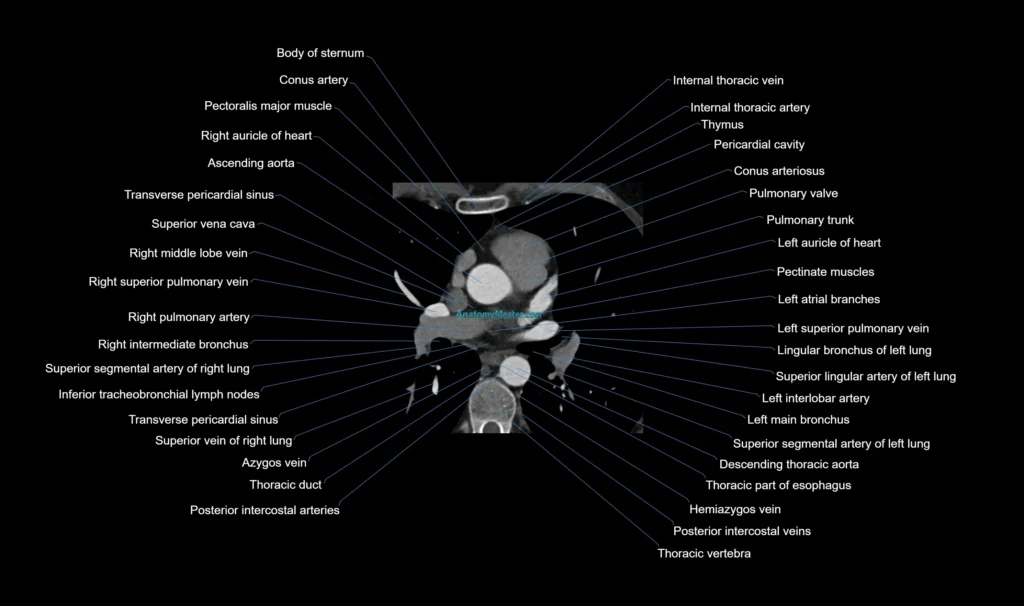
The axial CT images provide a systematic overview of the heart, coronary arteries, cardiac chambers, valves, pericardium and mediastinal structures. Users can scroll through the examination in the same way they would review a clinical CT coronary angiogram, helping them understand the changing appearance of cardiovascular anatomy from the cardiac base to the apex.
The two-chamber view demonstrates the left atrium, left ventricle, mitral valve, left ventricular apex and the anterior and inferior myocardial walls. This plane is particularly useful for understanding the relationship between the mitral valve, left ventricular cavity and long-axis myocardial anatomy.
The four-chamber view displays the right and left atria, right and left ventricles, interatrial septum, interventricular septum, mitral valve and tricuspid valve. It provides a clear comparison of the cardiac chambers and helps users understand atrioventricular anatomy, septal relationships and ventricular morphology.
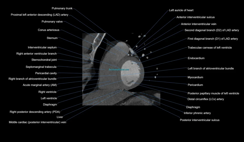
The short-axis view demonstrates the left and right ventricles in cross-section from the cardiac base to the apex. This plane is useful for studying the left ventricular myocardial segments, papillary muscles, ventricular cavities, interventricular septum and the relationship between the right and left ventricles.
The library also identifies important cardiac and vascular structures, including the ascending aorta, aortic root, sinuses of Valsalva, aortic valve, pulmonary trunk, right and left pulmonary arteries, pulmonary veins, superior vena cava, inferior vena cava, coronary sinus, left atrial appendage, right atrial appendage and pericardium.
Each CT coronary angiography image is manually labelled slice by slice in a textbook-style format, without the use of AI auto-labelling tools. This helps maintain anatomical accuracy and provides a reliable educational reference for radiologists, cardiologists, cardiac surgeons, radiology trainees, reporting radiographers, cardiac physiologists and students learning cardiovascular imaging.
The labelled images are displayed within a smooth interactive scroller, allowing users to move naturally through the anatomy as they would when reviewing a clinical cardiac CT examination. The platform works on desktop and mobile devices, while the full-screen viewer supports detailed examination of small coronary artery branches and complex cardiac structures.
The toolbar allows users to switch between axial, two-chamber, four-chamber and short-axis views. Specific structures can also be located using voice or text search, making it easier to identify coronary arteries, cardiac chambers, valves, myocardial segments, great vessels and other important anatomical landmarks.
The side zoom tool provides up to 10× magnification without loss of image quality, helping users examine the origins and courses of the coronary arteries, smaller arterial branches, valve anatomy and the relationships between cardiovascular structures in greater detail.
This module is supported by dynamic Level 1 and Level 2 quizzes that generate new questions during each session. These interactive assessments help users test their knowledge, revise cardiac and coronary artery anatomy and improve confidence when interpreting CT coronary angiography examinations.
CT Coronary Angiography Axial Anatomy: 1mm Labelled Image (Sample)
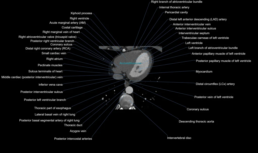
CT Coronary Angiography 2CH Anatomy: 1mm Labelled Image (Sample)
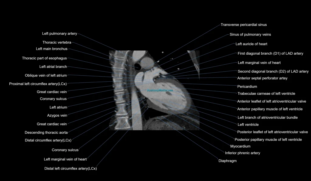
CT Coronary Angiography 4CH Anatomy: 1mm Labelled Image (Sample)
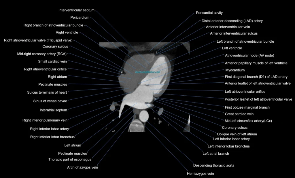
CT Coronary Angiography SA Anatomy: 1mm Labelled Image (Sample)