3T Temporomandibular Joint MRI Anatomy: Ultra-High-Resolution 1.5 mm Slices | Premium Subscription Access
Explore our advanced temporomandibular joint MRI anatomy library, featuring high-resolution labelled anatomical images of the TMJ and surrounding maxillofacial structures. This specialist learning resource is designed to demonstrate the mandibular condyle, articular disc, mandibular fossa, articular eminence, joint capsule, ligaments, and masticator muscles in a clinically realistic MRI format.
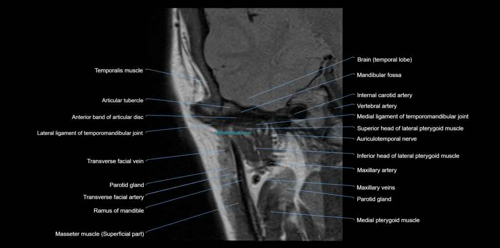
The TMJ MRI anatomy section includes detailed labelling of the mandibular condyle, glenoid fossa, articular eminence, articular disc, anterior band, posterior band, intermediate zone, retrodiscal tissue, bilaminar zone, joint capsule, lateral pterygoid muscle, medial pterygoid muscle, masseter muscle, temporalis muscle, mandibular ramus, zygomatic arch, temporal bone, and surrounding facial soft tissues.
Every individual image is manually labelled in a textbook-style format by our team, without the use of AI auto-labelling tools. This ensures high accuracy, consistent anatomical detail, and reliable learning support for radiologists, dentists, oral surgeons, maxillofacial surgeons, ENT surgeons, radiology students, dental students, reporting radiographers, and anyone interested in detailed TMJ anatomy.
Each labelled MRI image has been built into a smooth custom interactive scroller for easy learning. The interface is designed to be user-friendly on both desktop and mobile platforms. Double-clicking the scroller opens the image viewer in full-screen mode. The bottom toolbar allows users to switch between available imaging planes and use voice or text search to access detailed descriptions, including MRI and CT appearances of individual anatomical structures.
The side toolbar includes a powerful zoom function, allowing images to be magnified up to 10x without compromising image quality. This makes it easier to study the articular disc, condylar surface, retrodiscal tissue, joint space, capsule, and surrounding muscular attachments.
The labelling and anatomical content have been developed using multiple anatomy, dental, maxillofacial surgery, orthopaedic, and radiology reference books to provide a highly detailed and clinically focused learning experience. The work is being authorised and verified by experienced radiologists to ensure accuracy and reliability.
The temporomandibular joint MRI anatomy module is supported by Level 1 and Level 2 dynamic quizzes. These quizzes generate new questions in every session, helping users actively test their knowledge and build long-term understanding of TMJ anatomy.
This module is part of a separate radiology-focused anatomy website and is not available within the MRIMaster app. The full platform will be offered through an affordable annual subscription, with a strong focus on supporting students and healthcare professionals worldwide.
MRI Temporomandibular Joint sagittal 1.5 mm Deep Resolve Labelled Image(sample)
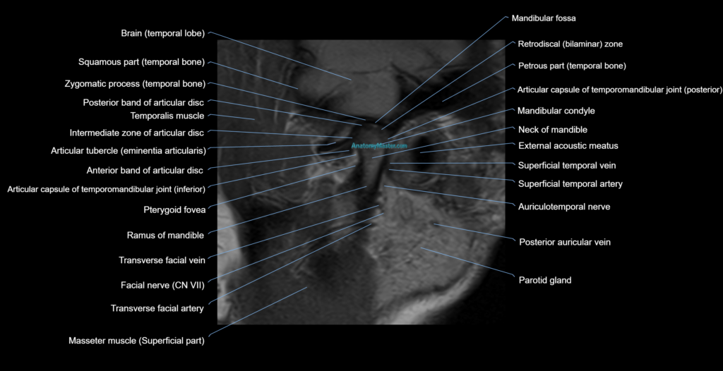
MRI Temporomandibular Joint coronal 1.5 mm Deep Resolve Labelled Image(sample)