3T Cranial Nerves MRI Anatomy Ultra-high-resolution 1.5mm Slices (Subscription-Based Premium Access)
Explore our advanced cranial nerves MRI anatomy library, featuring high-resolution labelled MRI images that demonstrate cranial nerves from their brainstem origins to their cisternal, cavernous sinus, skull base, orbital, and extracranial pathways. This module is designed for detailed learning of cranial nerve anatomy in axial, coronal, and sagittal planes.
The cranial nerve anatomy section includes the olfactory nerves, optic nerves, oculomotor nerves, trochlear nerves, trigeminal nerves, abducens nerves, facial nerves, vestibulocochlear nerves, glossopharyngeal nerves, vagus nerves, accessory nerves, and hypoglossal nerves. It also demonstrates key related structures such as the internal auditory meatus, cerebellopontine angle, Meckel’s cave, cavernous sinus, jugular foramen, hypoglossal canal, optic canal, and superior orbital fissure.
Each MRI slice has been manually labelled in a textbook-style format, without the use of AI auto-labelling tools. This is especially important for cranial nerve anatomy, where small structures can be difficult to identify and where precise anatomical relationships are clinically essential.
The interactive scroller allows users to follow cranial nerve pathways across multiple slices and planes. This helps build a practical understanding of nerve origins, courses, foramina, and relationships to vessels, brainstem structures, skull base bones, and surrounding soft tissues.
The bottom toolbar allows users to switch between axial, coronal, and sagittal planes and search for individual nerves or foramina using voice or text search. Detailed descriptions support learning of MRI and CT appearances of cranial nerve-related anatomy.
The 10x zoom tool helps users inspect fine structures such as the facial and vestibulocochlear nerves in the internal auditory canal, trigeminal nerve divisions, cavernous sinus nerves, and lower cranial nerves at the skull base.
The cranial nerves MRI anatomy module is supported by Level 1 and Level 2 quizzes that generate new questions each session, helping users actively revise and retain complex skull base and neuroanatomy.
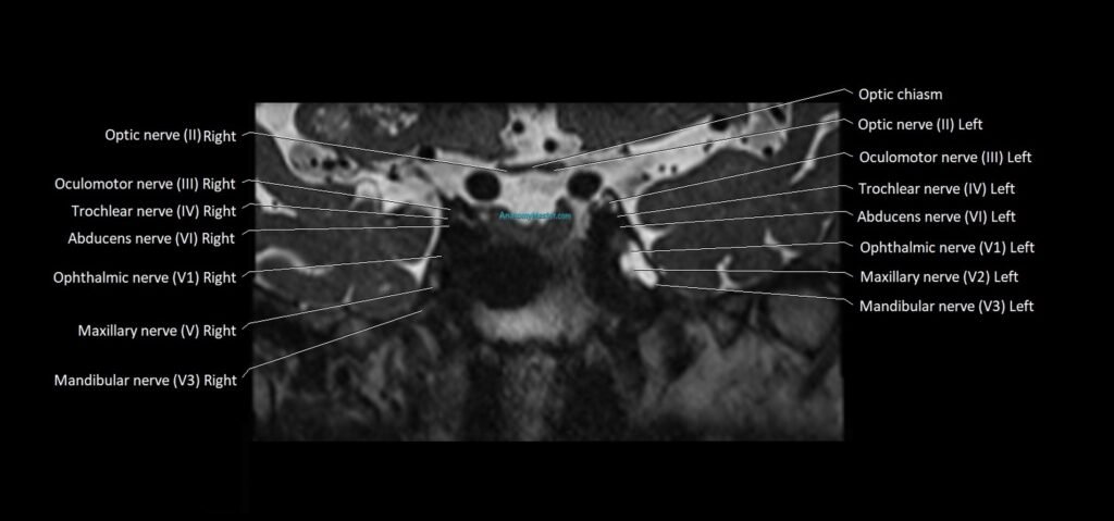
MRI Cranial Nerves Axial 1.5 mm Deep Resolve Labelled Image(sample)
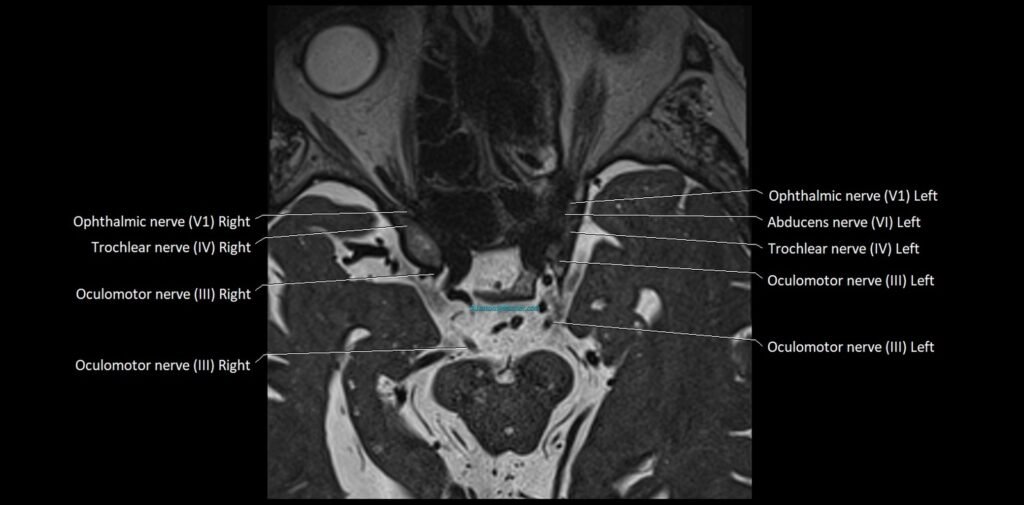
MRI Cranial Nerves sagittal 1.5 mm Deep Resolve Labelled Image(sample)
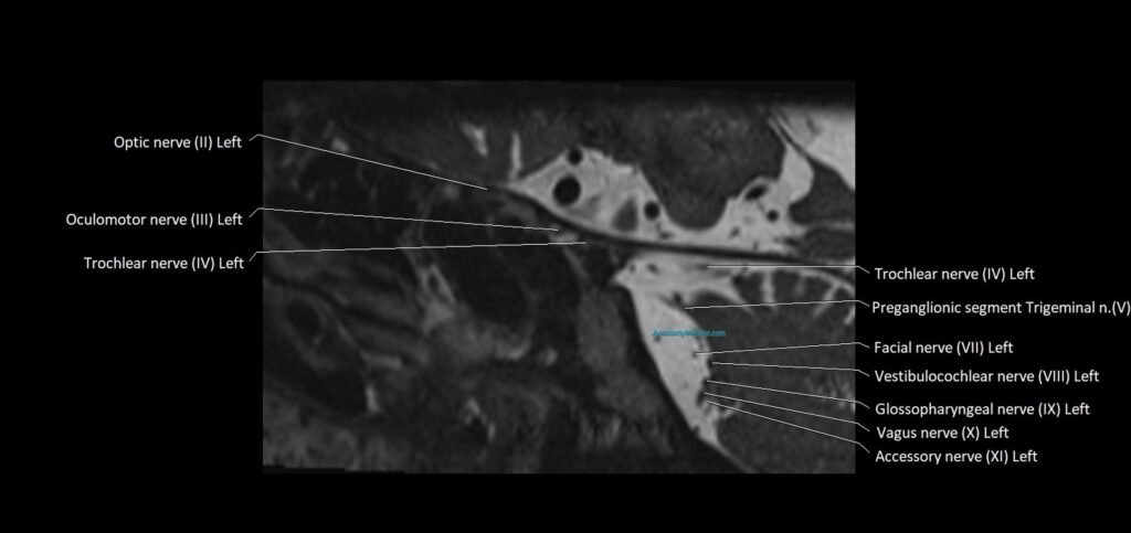
MRI Cranial Nerves coronal 1.5 mm Deep Resolve Labelled Image(sample)