3T Cerebellum MRI Anatomy Ultra-high-resolution 1.5mm Slices (Subscription-Based Premium Access)
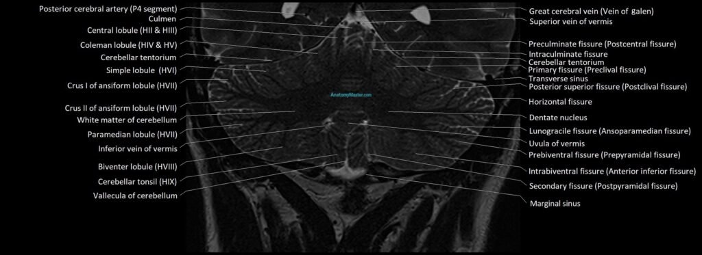
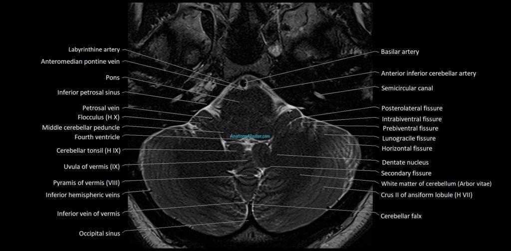
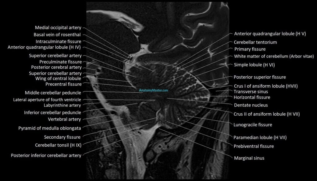
Explore our advanced cerebellum MRI anatomy library, featuring high-resolution labelled MRI images of the posterior fossa in axial, coronal, and sagittal planes. This specialist module is designed to help users understand the detailed anatomy of the cerebellar hemispheres, vermis, peduncles, nuclei, fourth ventricle, and surrounding cisternal spaces.
The cerebellum anatomy section includes the anterior lobe, posterior lobe, flocculonodular lobe, vermis, cerebellar tonsils, dentate nuclei, superior cerebellar peduncles, middle cerebellar peduncles, inferior cerebellar peduncles, cerebellopontine angles, fourth ventricle, brainstem relationships, and posterior fossa cisterns. These structures are essential for interpreting posterior fossa tumours, infarcts, Chiari malformation, cerebellar atrophy, vestibular pathway lesions, and surgical anatomy.
The MRI images are manually labelled in a clear textbook-style format, with each slice carefully reviewed for anatomical accuracy. No AI auto-labelling tools are used, allowing detailed and reliable labelling of small posterior fossa structures.
The interactive image scroller allows users to move smoothly through the cerebellum and posterior fossa, improving understanding of the three-dimensional relationship between the cerebellum, brainstem, ventricles, and skull base. The platform is designed for both desktop and mobile learning.
The bottom toolbar allows users to change between axial, coronal, and sagittal planes. Voice and text search help users quickly find cerebellar lobules, nuclei, peduncles, and related structures, with detailed descriptions to support imaging-based learning.
The side zoom function allows up to 10x magnification without compromising image quality, making it easier to study fine cerebellar anatomy and posterior fossa relationships.
The cerebellum MRI anatomy module is supported by dynamic quizzes, helping users test and retain knowledge of posterior fossa anatomy through active learning.
MRI Cerebellum Axial 1.5 mm Deep Resolve Labelled Image(sample)
MRI Cerebellum sagittal 1.5 mm Deep Resolve Labelled Image(sample)
MRI Cerebellum coronal 1.5 mm Deep Resolve Labelled Image(sample)