3T Hippocampus MRI Anatomy: Ultra-High-Resolution 1.5 mm Slices | Premium Subscription Access
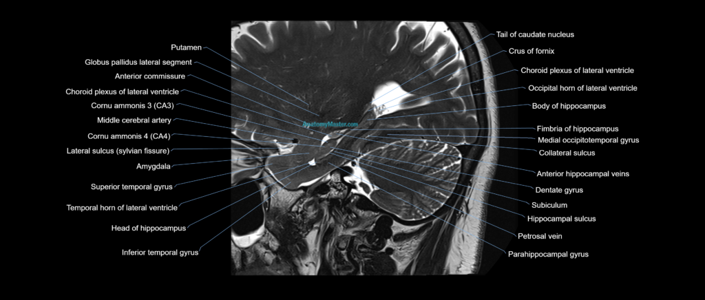
Explore our advanced hippocampus MRI anatomy library, featuring high-resolution labelled MRI images of the medial temporal lobe in coronal and sagittal planes. This module is designed to provide a detailed understanding of hippocampal anatomy and its relationship to surrounding limbic, ventricular, and temporal lobe structures.
The hippocampus anatomy section includes the hippocampal head, body and tail, pes hippocampi, dentate gyrus, subiculum, fimbria, fornix, amygdala, parahippocampal gyrus, temporal horn, choroid fissure, collateral sulcus, and surrounding medial temporal lobe anatomy. These structures are highly important in epilepsy imaging, dementia assessment, limbic system anatomy, and neurodegenerative disease interpretation.
The MRI images are selected for anatomical clarity and manually labelled slice by slice in a textbook-style format. No AI auto-labelling is used, ensuring that each anatomical label is carefully placed and checked for accuracy. This makes the module suitable for radiologists, neurologists, neurosurgeons, epilepsy teams, radiology trainees, and students learning detailed neuroanatomy.
The interactive image scroller allows users to follow the hippocampus through consecutive slices, helping them understand how the structure changes across different planes. The full-screen viewer provides a clear learning environment for studying small medial temporal lobe structures.
Users can switch between coronal and sagittal views using the bottom toolbar. Voice and text search allow quick access to individual anatomical structures, with detailed descriptions of their MRI and CT appearances where relevant.
The side zoom tool allows magnification up to 10x without compromising image quality. This is especially useful for evaluating fine hippocampal anatomy, internal architecture, and its relationship to the temporal horn and amygdala.
The hippocampus MRI anatomy module is supported by Level 1 and Level 2 dynamic quizzes, which generate new questions every session to reinforce active learning and long-term anatomical knowledge.
MRI Cavernous Sinus coronal 1.5 mm Deep Resolve Labelled Image(sample)
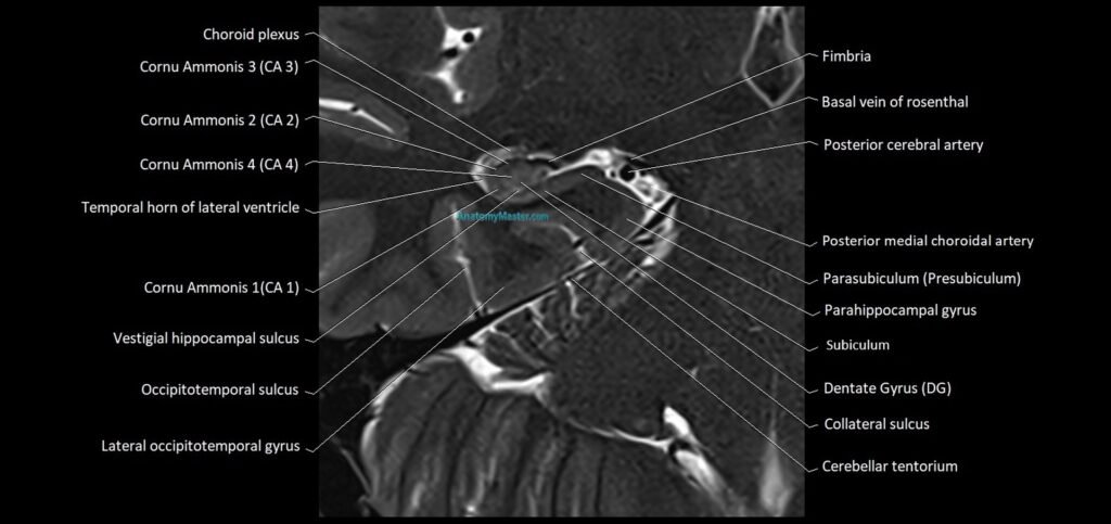
MRI Cavernous Sinus sagittal 1.5 mm Deep Resolve Labelled Image(sample)