3T Cavernous Sinus MRI Anatomy: Ultra-High-Resolution 1.5 mm Slices | Premium Subscription Access
Explore our advanced cavernous sinus MRI anatomy library, featuring high-resolution labelled MRI images in axial and coronal planes. This specialist module is designed to help users understand one of the most complex regions of skull base anatomy, where cranial nerves, venous channels, the internal carotid artery, pituitary region, and orbital apex structures are closely related.
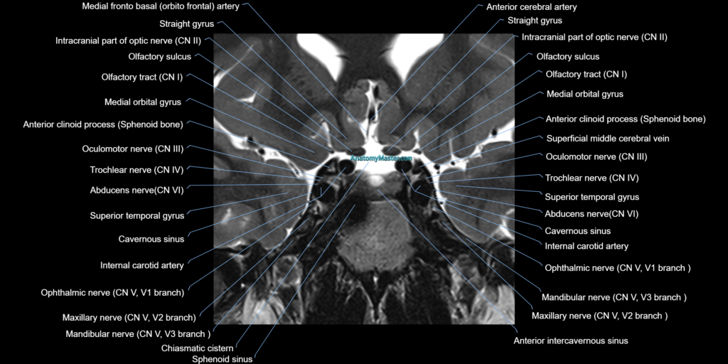
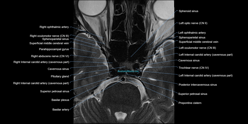
The cavernous sinus anatomy section demonstrates the internal carotid artery, oculomotor nerve, trochlear nerve, ophthalmic nerve, maxillary nerve, abducens nerve, pituitary gland, sphenoid sinus, Meckel’s cave, superior orbital fissure, optic canal, and surrounding skull base landmarks. These relationships are essential for interpreting cavernous sinus lesions, pituitary extension, cranial neuropathies, vascular abnormalities, infection, inflammation, and skull base tumours.
The images are manually labelled slice by slice in a clear textbook-style format, without AI auto-labelling. This allows accurate identification of small structures that are often difficult to appreciate on standard MRI. The module is particularly useful for radiologists, ENT surgeons, neurosurgeons, ophthalmologists, radiology trainees, and reporting radiographers.
Each labelled MRI image is integrated into a smooth custom scroller, allowing the user to move through the cavernous sinus region in the same way as a clinical MRI study. The interface is simple to use on both desktop and mobile devices, with a full-screen option for detailed review.
The bottom toolbar allows users to switch between axial and coronal planes and search for specific anatomical structures using voice or text search. Detailed descriptions help explain the MRI and CT appearance of key structures in and around the cavernous sinus.
The 10x zoom function allows close inspection of cranial nerves, vascular channels, and skull base foramina without compromising image quality. This is especially important for understanding the small but clinically significant anatomy of the cavernous sinus.
The module is supported by dynamic anatomy quizzes, helping users test their knowledge and improve long-term recall of cavernous sinus and skull base anatomy.
MRI Cavernous Sinus Axial 1.5 mm Deep Resolve Labelled Image(sample)
MRI Cavernous Sinus coronal 1.5 mm Deep Resolve Labelled Image(sample)