3T Brainstem and Basal Ganglia MRI Anatomy: Ultra-High-Resolution 1.5 mm Slices | Premium Subscription Access
Explore our advanced brainstem and basal ganglia MRI anatomy library, featuring ultra-high-resolution anatomical images in axial, sagittal, and coronal planes. This specialist learning module has been developed from repeated volunteer MRI scans using advanced 3T scanners, dedicated high-channel coils, and Deep Resolve image enhancement to demonstrate clinically realistic neuroanatomy.
The brainstem and basal ganglia section focuses on complex deep brain structures, including the midbrain, pons, medulla, thalamus, caudate nucleus, lentiform nucleus, internal capsule, external capsule, corticospinal tracts, cerebellar peduncles, cranial nerve nuclei, and surrounding vascular and ventricular landmarks. These areas are often difficult to understand on routine imaging, so the module is designed to make deep neuroanatomy clearer and easier to learn.
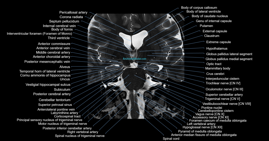
The MRI images are manually labelled slice by slice in a textbook-style format, without the use of AI auto-labelling tools. This helps preserve anatomical accuracy and provides a reliable learning reference for radiologists, neurologists, neurosurgeons, radiology trainees, reporting radiographers, and students preparing for neuroimaging interpretation.
Each labelled image is displayed within a smooth interactive scroller, allowing users to move through the anatomy naturally as they would review a clinical MRI study. The platform works on desktop and mobile devices, and the full-screen viewer allows detailed review of small anatomical structures.
The toolbar allows users to move between axial, coronal, and sagittal planes and search for specific structures using voice or text search. This helps users quickly locate important structures such as the substantia nigra, red nucleus, cerebral peduncles, internal capsule, basal ganglia nuclei, and brainstem cranial nerve pathways.
The side zoom tool allows up to 10x magnification without loss of image quality, helping users study fine internal brainstem anatomy and deep grey matter relationships in greater detail.
This module is supported by dynamic Level 1 and Level 2 quizzes that generate new questions in every session, helping users actively revise and retain complex brainstem and basal ganglia anatomy.
MRI Brainstem Axial 1.5 mm Deep Resolve Labelled Image(sample)
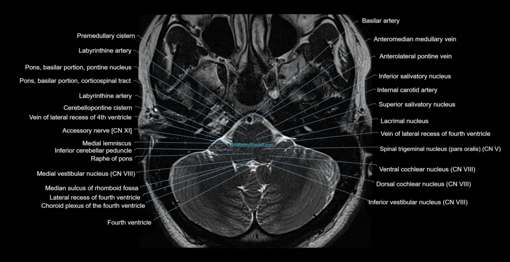
MRI Brainstem sagittal 1.5 mm Deep Resolve Labelled Image(sample)
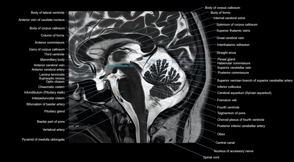
MRI Brainstem coronal 1.5 mm Deep Resolve Labelled Image(sample)