Spondylolisthesis MRI
Spondylolisthesis is a spinal condition wherein one vertebra shifts forward or backward in relation to the one below it. This displacement can result in a range of symptoms and potential complications. Although it can occur anywhere along the spine, it is most frequently observed in the lower back. The causes, symptoms, and treatment options differ depending on the type and severity of the condition.
Causes
Spondylolisthesis can be caused by several factors:
- Congenital Factors: Some individuals are born with a predisposition due to the abnormal bone formation of the spine.
- Degenerative Disease: Often seen in adults due to aging-related wear and tear on the spine, especially in the intervertebral discs and joints.
- Traumatic Causes: An injury or trauma to the spine, such as from sports, accidents, or severe impact, can lead to spondylolisthesis.
- Pathological Causes: Conditions like osteoporosis (weakening of bones) or tumor growth can weaken the spine, leading to slippage.
- Isthmic Spondylolisthesis: Occurs due to a small stress fracture, usually in adolescents involved in athletic activities.
Symptoms
Symptoms vary but may include:
- Lower back pain, especially after prolonged standing or walking.
- Stiffness or tightness in the back muscles.
- Pain radiating down the legs (sciatica), often due to nerve compression.
- Numbness, tingling, or weakness in the lower extremities.
- Reduced range of motion in the lower back.
- In severe cases, changes in posture or gait.
Treatment
Treatment for spondylolisthesis varies based on the severity and type of the condition:
Non-Surgical Treatment:
- Physical Therapy: To strengthen the muscles in the back and abdomen.
- Medications: Pain relievers and anti-inflammatory drugs.
- Activity Modification: Avoid activities that strain the back.
- Bracing: In some cases, wearing a back brace can help.
Surgical Treatment:
Recommended in severe cases, especially when conservative treatments fail to relieve symptoms.
- Spinal Fusion: The most common surgery, which involves fusing the affected vertebrae to prevent them from slipping.
MRI Appearance of Spondylolisthesis
- T1-weighted MRI: On a T1-weighted MRI, normal bone marrow is usually high in signal intensity, appearing bright. In the case of spondylolisthesis, the slipped vertebra can be clearly identified. The alignment of the vertebrae is easily observable, and any associated changes in the bone marrow can be detected. For instance, if there is marrow edema or inflammation, it may appear as a lower signal area within the bright bone marrow.
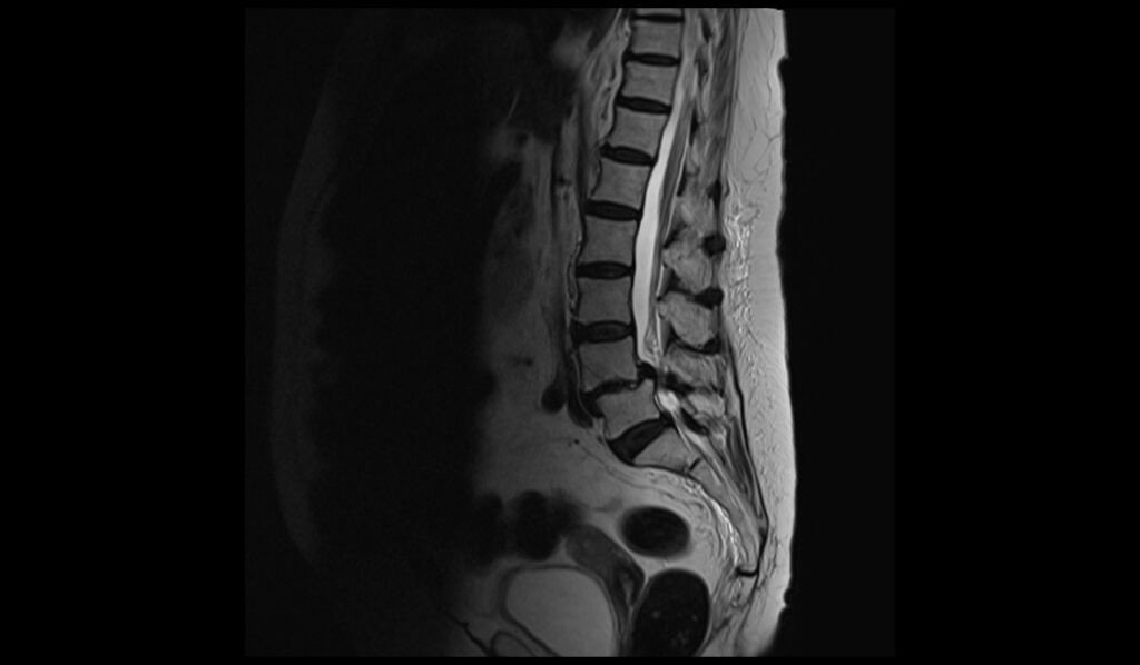
- T2-weighted MRI: This sequence provides images where water content appears bright. It is especially useful for evaluating the condition of soft tissues, including discs, ligaments, and nerves. In the context of spondylolisthesis, T2-weighted images can help in assessing the degree of nerve root compression, if present, and the status of the intervertebral discs. It also aids in evaluating any associated conditions such as spinal stenosis or disc herniation.
- STIR (Short Tau Inversion Recovery): STIR is a special type of MRI sequence that is used to suppress the signals from fat. This makes it highly sensitive to changes in water content, thus highlighting areas of inflammation or edema (swelling). In spondylolisthesis, STIR can be particularly useful in identifying bone marrow edema, which might suggest an acute injury or stress reaction in the bone.
T2 sagittal image shows spondylolisthesis of lumbar spine
STIR sagittal image shows spondylolisthesis of lumbar spine
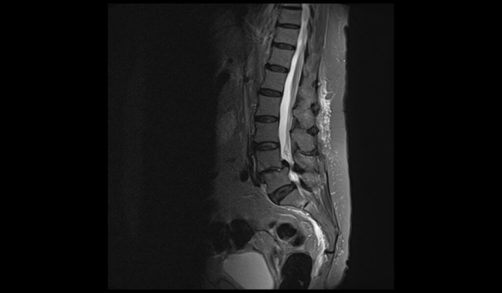
T1 TSE sagittal image shows spondylolisthesis of lumbar spine

T2 TSE axial image shows spondylolisthesis of lumbar spine

T1 TSE axial image shows spondylolisthesis of lumbar spine

References
- Li, N., Scofield, J., Mangham, P., Cooper, J., Sherman, W., & Kaye, A. D. (2022). Spondylolisthesis. Orthop Rev (Pavia), 14(3), 36917. doi: 10.52965/001c.36917. PMID: 35910544. PMCID: PMC9329062.
- de Dios, E., Laesser, M., Björkman-Burtscher, I. M., Lindhagen, L., & MacDowall, A. (2023). MRI-based measurements of spondylolisthesis and kyphosis in degenerative cervical myelopathy. BMC Medical Imaging, 23, 180. doi:10.1186/s12880-023-00829-5
- Cho, I. Y., Park, S. Y., & Lee, S. H. (2017). MRI findings of lumbar spine instability in degenerative spondylolisthesis. Journal of Orthopaedic Surgery, 1(1), 1-10.
- Nakayama, T., & Ehara, S. (2015). Spondylolytic spondylolisthesis: various imaging features and natural courses. Japanese Journal of Radiology, 33(1), 3–12. Published on 23 November 2014.