MRI Thigh Two-Station Automatic Composing Protocols and Planning
Automatic Composing in MRI
Automatic composing in MRI refers to a technique used to create a continuous, seamless image from multiple adjacent scans. This is particularly necessary for imaging long structures such as the spine or long bones, where a single scan may not cover the entire area of interest. In large-diameter and short-bore MRI scanners, the field of view (FOV) is limited, making it challenging to capture long anatomical regions in a single scan.
With automatic composing, the MRI scanner acquires images in overlapping segments or stations. These individual images are then automatically stitched together by the software to form a comprehensive view of the entire area. This technique ensures that detailed and accurate images are obtained without gaps or misalignments, providing a complete diagnostic picture. Automatic composing is essential for accurate diagnosis and treatment planning, particularly in conditions that affect long sections of the spine or large bones.
Indications for mri thigh scan
- Marrow abnormalities: avascular necrosis, marrow edema syndromes, and stress fractures
- Muscle and tendon disorders: strains, partial and complete tears, tendonitis, tendonopathy
- Congenital and developmental conditions
- Neoplasms of bone, joint or soft tissue
- Infections of bone, joint or soft tissue
- Myositis and osteomyelitis
- Hamstring injury
- Ligament tears
- Acute trauma
- fractures
- Abcess
- Ulcer
Contraindications
- Any electrically, magnetically or mechanically activated implant (e.g. cardiac pacemaker, insulin pump biostimulator, neurostimulator, cochlear implant, and hearing aids)
- Intracranial aneurysm clips (unless made of titanium)
- Pregnancy (risk vs benefit ratio to be assessed)
- Ferromagnetic surgical clips or staples
- Metallic foreign body in the eye
- Metal shrapnel or bullet
Patient preparation
- A satisfactory written consent form must be taken from the patient before entering the scanner room
- Ask the patient to remove all metal object including keys, coins, wallet, any cards with magnetic strips, jewellery, hearing aid and hairpins
- If possible offer some one in the scanner room for claustrophobic patient (e.g. relative or staff ).
- Offer earplug or headphones possibly with music for extra comfort.
- Properly explain the procedure to the patient
- Instruct the patient to keep still
- Note the weight of the patient
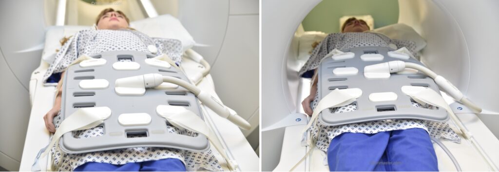
Positioning for mri upper legs
- Position the patient in supine position with feet pointing towards the magnet (feet first supine)
- Position the patient over the spine coil and place the body coils over the thighs (anterior superior iliac spine down to knee joints)
- Securely tighten the body coil using straps
- Centre the laser beam localizer over the mid thigh
Recommended MRI thigh Automatic Composing Protocols and Planning
LOCALIZER THIGH AUTOMATIC COMPOSING
The auto-composing localizer system consists of two distinct localizers: one for the upper thigh and another for the lower thigh. After completing the localizer for the upper thigh, the table will automatically reposition itself to perform the lower thigh localizer. Once both localizers are finished, the system will merge them together to generate a coronal and sagittal localizer for the entire thigh.

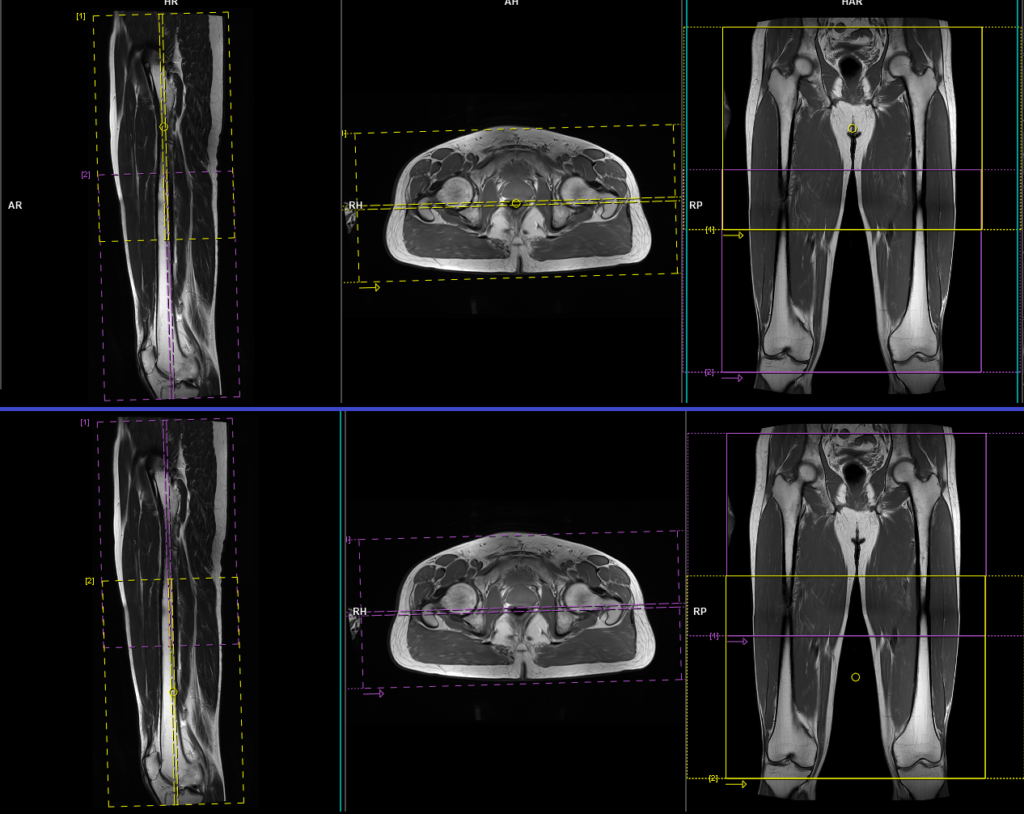
T2 stir coronal 5mm AUTOMATIC COMPOSING
When using the auto-composing protocol, both the upper thigh and lower thigh blocks will appear on the thigh auto-compose localizer. The planning should begin with the upper thigh block, and once it is fully completed, move on to the lower thigh block. It is important to ensure sufficient overlap between the blocks. Once both blocks are planned with proper positioning and alignment, the user can initiate the scan. After both scans are completed, the system will automatically compose the blocks and generate whole-thigh sagittal images.
For the upper thigh block, it should be planned on the sagittal plane, and the positioning block should be angled parallel to the femur. It is necessary to check the positioning block in the axial and coronal planes. In the axial plane, the angle should be parallel to the right and left femur. In the coronal plane, the field of view (FOV) should cover the upper thigh from 2 inches above the hip joint down to 1 inch below the mid-thigh, typically ranging from 300-350 mm with a phase FOV of 120%. The slices must be sufficient to cover the entire thigh from anterior to posterior.
Similarly, for the lower thigh block, it should be planned on the sagittal plane, and the positioning block should be angled parallel to the femur. It is necessary to check the positioning block in the axial and coronal planes. In the axial plane, the angle should be parallel to the right and left femur. In the coronal plane, the FOV should cover the lower thigh from 2 inches above the mid-thigh down to 2 inches below the knee joint, typically ranging from 300-350 mm with a phase FOV of 120%. The slices must be sufficient to cover the entire thigh from anterior to posterior.
Parameters
TR 4000-5000 | TE 110 | FLIP 150 | NEX 2 | SLICE 5 MM | MATRIX 384X320 | FOV 300-350 | PHASE R>L | GAP 10% | TI 150 |
T1 tse coronal 5mm AUTOMATIC COMPOSING
Plan the upper thigh block on the sagittal plane, and angle the positioning block parallel to the femur. It is necessary to check the positioning block in both the axial and coronal planes. In the axial plane, the angle should be parallel to the right and left femur. In the coronal plane, the field of view (FOV) should cover the upper thigh from 2 inches above the hip joint down to 1 inch below the mid-thigh, typically ranging from 300-350 mm with a phase FOV of 120%. The slices must be sufficient to cover the entire thigh from anterior to posterior.
Similarly, for the lower thigh block, plan it on the sagittal plane and angle the positioning block parallel to the femur. It is necessary to check the positioning block in both the axial and coronal planes. In the axial plane, the angle should be parallel to the right and left femur. In the coronal plane, the FOV should cover the lower thigh from 2 inches above the mid-thigh down to 2 inches below the knee joint, typically ranging from 300-350 mm with a phase FOV of 120%. The slices must be sufficient to cover the entire thigh from anterior to posterior.
Parameters
TR 400-600 | TE 15-25 | SLICE 5 MM | FLIP 160 | PHASE R>L | MATRIX 448X448 | FOV 300-350 | GAP 10% | NEX(AVRAGE) 2 |
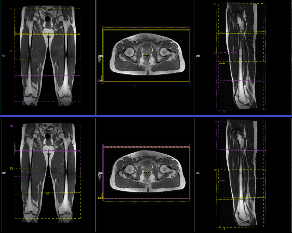
T1 tse axial 6mm AUTOMATIC COMPOSING
For the upper thigh block, it should be planned on the sagittal plane, and the positioning block should be angled perpendicular to the femur. It is necessary to check the positioning block in the coronal and axial planes. In the coronal plane, the angle should be perpendicular to the femur. In the axial plane, the field of view (FOV) should cover the entire thigh, typically ranging from 300-350 mm with a phase FOV of 120%. The slices must be sufficient to cover the upper thigh from 2 inches above the hip joint down to 1 inch below the mid-thigh.
Similarly, for the lower thigh planning block, it should be planned on the sagittal plane, and the positioning block should be angled perpendicular to the femur. It is necessary to check the positioning block in the coronal and axial planes. In the coronal plane, the angle should be perpendicular to the right and left femur. In the axial plane, the FOV should cover the entire thigh, typically ranging from 300-350 mm with a phase FOV of 120%. The slices must be sufficient to cover the lower thigh from 2 inches above the mid-thigh down to 2 inches below the knee joint.
Parameters
TR 400-600 | TE 15-25 | SLICE 6 MM | FLIP 160 | PHASE A>P | MATRIX 448X448 | FOV 300-350 | GAP 10% | NEX(AVRAGE) 2 |
T2 stir axial 6mm AUTOMATIC COMPOSING
For the upper thigh block, it should be planned on the sagittal plane, and the positioning block should be angled perpendicular to the femur. It is necessary to check the positioning block in the coronal and axial planes. In the coronal plane, the angle should be perpendicular to the femur. In the axial plane, the field of view (FOV) should cover the entire thigh, typically ranging from 300-350 mm with a phase FOV of 120%. The slices must be sufficient to cover the upper thigh from 2 inches above the hip joint down to 1 inch below the mid-thigh.
Similarly, for the lower thigh planning block, it should be planned on the sagittal plane, and the positioning block should be angled perpendicular to the femur. It is necessary to check the positioning block in the coronal and axial planes. In the coronal plane, the angle should be perpendicular to the right and left femur. In the axial plane, the FOV should cover the entire thigh, typically ranging from 300-350 mm with a phase FOV of 120%. The slices must be sufficient to cover the lower thigh from 2 inches above the mid-thigh down to 2 inches below the knee joint.
Parameters
TR 4000-5000 | TE 110 | FLIP 160 | NEX 2 | SLICE 6 MM | MATRIX 384X320 | FOV 300-350 | PHASE A>P | GAP 10% | TI 150 |
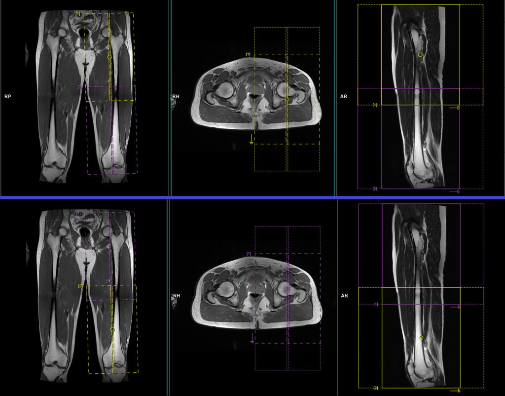
T2 tse sagittal 5mm AUTOMATIC COMPOSING
Plan the upper thigh block on the coronal plane, angling the positioning block parallel to the femoral shaft. Check the positioning block in the other two planes. An appropriate angle must be given in the axial plane (vertically across the left thigh). In the coronal plane, the field of view (FOV) should cover the upper thigh from 2 inches above the hip joint down to 1 inch below the mid-thigh, typically ranging from 300-350 mm with a phase FOV of 120%. The slices must be sufficient to cover the left thigh from medial to lateral.
Similarly, for the lower thigh planning block, it should be planned on the coronal plane, with the positioning block angled parallel to the femoral shaft. Check the positioning block in the other two planes. An appropriate angle must be given in the axial plane (vertically across the left thigh). In the coronal plane, the FOV should cover the lower thigh from 2 inches above the mid-thigh down to 2 inches below the knee joint, typically ranging from 300-350 mm with a phase FOV of 120%. The slices must be sufficient to cover the left thigh from medial to lateral.
Parameters
TR 4000-5000 | TE 100-120 | SLICE 5 MM | FLIP 130-150 | PHASE A>P | MATRIX 384X384 | FOV 300-350 | GAP 10% | NEX(AVRAGE) 2 |