Temporomandibular Joint (TMJ) MRI
Introduction
Magnetic resonance imaging (MRI) is the most valuable and extensively utilized imaging technique for assessing the temporomandibular joint (TMJ). In comparison to computed tomography (CT) and arthrography, MRI offers superior tissue contrast, enabling clear visualization of soft tissues and other articular structures within the TMJ. Notably, MRI allows for direct visualization of the articular disc without the need for any contrast medium, giving it an edge over arthrography. The proper selection of coils and meticulous imaging techniques significantly contribute to successful TMJ imaging.
TMJ Anatomy
The temporomandibular joint (TMJ) is a crucial and complex anatomical structure that plays a central role in jaw movement and function. It is a bilateral joint located on either side of the head, connecting the lower jaw (mandible) to the temporal bone of the skull. The TMJ is responsible for facilitating essential functions like chewing, speaking, and swallowing.
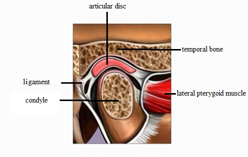
Anatomically, the TMJ is comprised of several key components that work together harmoniously. The articular surfaces of the joint consist of the rounded condyle of the mandible and the concave mandibular fossa on the temporal bone. Separating these surfaces is a fibrocartilaginous articular disc, which acts as a cushion and helps to distribute forces during jaw movements.
Ligaments surrounding the TMJ provide stability and limit excessive movements. The lateral ligament, also known as the temporomandibular ligament, is the primary stabilizer and prevents the mandible from moving too far backward. The stylomandibular ligament and the sphenomandibular ligament also contribute to joint stability.
Muscles surrounding the TMJ are vital for its proper functioning. The muscles of mastication, including the temporalis, masseter, medial pterygoid, and lateral pterygoid muscles, are responsible for jaw movements during chewing. Disorders of the TMJ can lead to various symptoms such as pain, clicking, or difficulty in jaw movement.
Please check our new video tutorial for protocols and planning
Indications for TMJ MRI Scan
- Irregular jaw movement with difficulty in opening and closing the mouth
- Pain in the ear area when speaking, chewing or opening the mouth wide
- TMJ Headaches (become worse while opening and closing the mouth)
- Clicking sounds in the jaw joint when opening or closing the mouth
- Ear pain in front of or below the ear without any signs of infection
- Pain in the jaw joint area, tooth, neck and shoulders
- Jaw pain or toothache when waking up after sleep
- Feelings of teeth are not fitting together properly
- Difficulty in chewing
Contraindications
- Any electrically, magnetically or mechanically activated implant (e.g. cardiac pacemaker, insulin pump biostimulator, neurostimulator, cochlear implant, and hearing aids)
- Intracranial aneurysm clips (unless made of titanium)
- Pregnancy (risk vs benefit ratio to be assessed)
- Ferromagnetic surgical clips or staples
- Metallic foreign body in the eye
- Metal shrapnel or bullet
Patient preparation for TMJ MRI scan
- A satisfactory written consent form must be taken from the patient before entering the scanner room
- Ask the patient to remove all metal objects including keys, coins, wallet, cards with magnetic strips, jewellery, hearing aid and hairpins
- Ask the patient to undress and change into a hospital gown
- Instruct the patient keep still explain it is a long procedure (give an estimation of time so that they can mentally prepare themselves )
- If possible provide a chaperone for claustrophobic patients (e.g. relative or staff )
- Offer earplugs or headphones, possibly with music for extra comfort
- Note the hight and weight of the patient
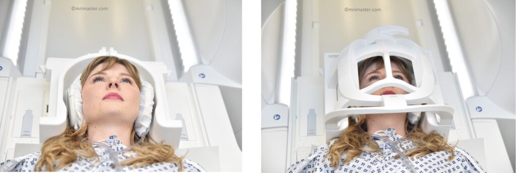
Positioning for TMJ MRI Scan
- Head first supine
- Position the head in the head coil and immobilise with cushions
- Give cushions under the legs for extra comfort
- Centre the laser beam localizer over the glabella
Recommended TMJ MRI Protocols, Parameters, and Planning
TMJ MRI Localizer
To plan the sequences, it is essential to acquire a three-plane localizer. These localizers typically have a duration of less than 25 seconds and are T1-weighted with low resolution.

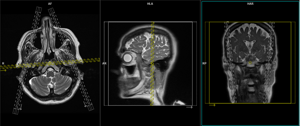
T2 tse axial
Plan the axial slices on the sagittal plane and angle the planning block parallel to the hard palate. The slices must be sufficient to cover the mandible from the corpus callosum up to the line of the angle of the jaw. Check the positioning block in the other two planes, ensuring an appropriate angle is given in the coronal plane (perpendicular to the brain stem)
Parameters

TR 3000-4000 | TE 100-120 | SLICE 4MM | FLIP 130-150 | PHASE R>L | MATRIX 320X320 | FOV 210-230 | GAP 10% | NEX(AVRAGE) 2 |
TMJ scan planning localizers
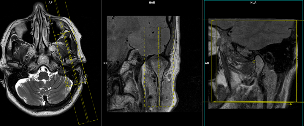
Plan the sagittal localizers on the axial plane: for the right sagittal localizer, angle the planning block perpendicular to the right condyle of the mandible, and for the left side localizer, angle the planning block perpendicular to the left condyle of the mandible. Check the planning block in the other two planes. An appropriate angle must be given in the coronal plane (parallel to the line along the temporal bone and ramus of the mandible).
Plan the coronal localizer on the axial plane: angle the planning block parallel to the line along the left and right mandibular condyles. Check the positioning block in the sagittal plane (parallel to the mandibular ramus).
PD coronal 2mm SFOV RT TMJ
Plan the right side coronal slices on the axial plane, and angle the planning block parallel to the right condyle of the mandible. Check the planning block in the other two planes. An appropriate angle must be given in the sagittal plane (parallel to the line along the ramus and right mandibular condyle). Ensure that the slices are sufficient to cover the right temporomandibular joint (RT TMJ) from the articular eminence up to the line of the internal auditory meatus.
Parameters
TR 2500-3500 | TE 15-30 | FLIP 150 | NEX 2 | SLICE 2MM | MATRIX 256×256 | FOV 100 | PHASE R>L | OVERSAMPLE 80% | FAT SAT NO |
PD coronal 2mm SFOV LT TMJ
Plan the left side coronal slices on the axial plane; angle the planning block parallel to the left condyle of the mandible. Check the planning block in the other two planes. Ensure an appropriate angle is given in the sagittal plane, parallel to the line along the ramus and left mandibular condyle. The slices must be sufficient to cover the left temporomandibular joint (TMJ) from the articular eminence up to the line of the internal auditory meatus.
Parameters

TR 2500-3500 | TE 15-30 | FLIP 150 | NEX 2 | SLICE 2MM | MATRIX 256×256 | FOV 100 | PHASE R>L | OVERSAMPLE 80% | FAT SAT NO |
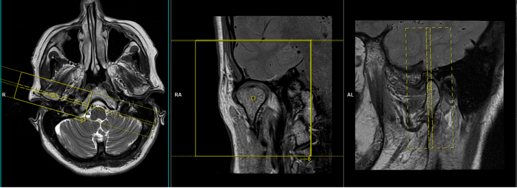
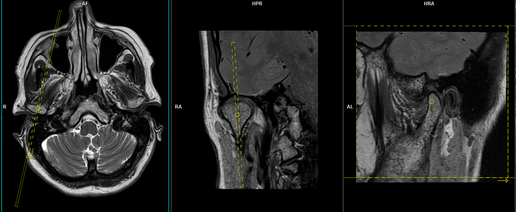
PD sagittal 2mm SFOV RT TMJ Close mouth
Plan the right side sagittal slices on the axial plane; angle the planning block perpendicular to the right condyle of the mandible. Check the planning block in the other two planes. An appropriate angle must be given in the coronal plane (parallel to the line along the right temporal bone and ramus of the mandible). The slices should adequately cover the right temporomandibular joint (TMJ) from one side to the other.

Parameters
TR 2500-3500 | TE 15-30 | FLIP 150 | NEX 2 | SLICE 2MM | MATRIX 256×256 | FOV 100 | PHASE R>L | OVERSAMPLE 80% | FAT SAT NO |
PD sagittal 2mm SFOV LT TMJ Close mouth
Plan the left side sagittal slices on the axial plane; angle the planning block perpendicular to the left condyle of the mandible. Check the planning block in the other two planes. An appropriate angle must be given in the coronal plane (parallel to the line along the left temporal bone and ramus of the mandible). The slices should adequately cover the left temporomandibular joint (TMJ) from one side to the other.
Parameters
TR 2500-3500 | TE 15-30 | FLIP 150 | NEX 2 | SLICE 2MM | MATRIX 256×256 | FOV 100 | PHASE R>L | OVERSAMPLE 80% | FAT SAT NO |
STIR sagittal 2mm SFOV RT TMJ Close mouth
Plan the right side sagittal slices on the axial plane; angle the planning block perpendicular to the right condyle of the mandible. Check the planning block in the other two planes. An appropriate angle must be given in the coronal plane (parallel to the line along the right temporal bone and ramus of the mandible). The slices should adequately cover the right temporomandibular joint (TMJ) from one side to the other.
Parameters
TR 3000-4000 | TE 90-100 | FLIP 130 | NEX 3 | SLICE 2MM | MATRIX 256×224 | FOV 100 | PHASE A>P | OVERSAMPLE 100% | TI 150 |
STIR sagittal 2mm SFOV LT TMJ Close mouth
Plan the left side sagittal slices on the axial plane; angle the planning block perpendicular to the left condyle of the mandible. Check the planning block in the other two planes. An appropriate angle must be given in the coronal plane (parallel to the line along the left temporal bone and ramus of the mandible). The slices should adequately cover the left temporomandibular joint (TMJ) from one side to the other.
Parameters
TR 3000-4000 | TE 90-100 | FLIP 130 | NEX 3 | SLICE 2MM | MATRIX 256×224 | FOV 100 | PHASE A>P | OVERSAMPLE 100% | T1 150 |
PD sagittal 2mm SFOV RT TMJ open mouth
Plan the right side open mouth sagittal slices on the axial plane; angle the planning block perpendicular to the right condyle of the mandible. Check the planning block in the other two planes. An appropriate angle must be given in the coronal plane (parallel to the line along the right temporal bone and ramus of the mandible). The slices should adequately cover the right temporomandibular joint (TMJ) from one side to the other.

Parameters
TR 2500-3500 | TE 15-30 | FLIP 150 | NEX 2 | SLICE 2MM | MATRIX 256×256 | FOV 100 | PHASE R>L | OVERSAMPLE 80% | FAT SAT NO |
PD sagittal 2mm SFOV LT TMJ open mouth
Plan the left side open mouth sagittal slices on the axial plane; angle the planning block perpendicular to the left condyle of the mandible. Check the planning block in the other two planes. An appropriate angle must be given in the coronal plane (parallel to the line along the left temporal bone and ramus of the mandible). The slices should adequately cover the left temporomandibular joint (TMJ) from one side to the other.

Parameters
TR 2500-3500 | TE 15-30 | FLIP 150 | NEX 2 | SLICE 2MM | MATRIX 256×256 | FOV 100 | PHASE R>L | OVERSAMPLE 80% | FAT SAT NO |
Optional Scans
TRUFISP\FLASH CINE DYNAMIC sagittal 3mm RT
A small FOV True Fast Imaging with Steady Precession (TRUFISP) cine scan can provide dynamic real-time movement information of the TMJ (Temporomandibular Joint). This imaging technique is commonly used as a supplementary scan to gather additional information about TMJ anomalies during motion, such as dislocation. To conduct a dynamic scan, a single-slice TRUFISP sequence is used, lasting approximately 1 minute. During this time, the patient is instructed to slowly open and close their mouth to observe the joint in motion. Since this sequence is employed to gather supplementary information, a fast 1-minute dynamic scan is generally sufficient.
Some other radiologists prefer using single-slice high-resolution PD or T1 imaging in the same position but with different mouth opening positions, utilizing various bite blocks. The images are then combined to create dynamic sequences. However, this technique requires a minimum of 10 minutes for each side. In our department, we prefer the first method to save time.
Plan the right side dynamic sagittal slice on the axial plane; angle the planning block perpendicular to the right condyle of the mandible. Check the planning block in the other two planes. An appropriate angle must be given in the coronal plane (parallel to the line along the right temporal bone and ramus of the mandible).
Parameters
TR 4-5 | TE 2-3 | FLIP 60 | NEX 1 | SLICE 3 MM | MATRIX 256×256 | FOV 150-200 | PHASE A>P | OVERSAMPLE 50% | IPAT OFF |

