MRI Spinal epidural hematoma
A spinal epidural hematoma (SEH) is a collection of blood that accumulates in the epidural space, which is the area between the outer covering of the spinal cord (dura mater) and the vertebrae. This condition can compress the spinal cord and nerves, leading to potentially serious neurological deficits.
Causes
Spinal epidural hematomas can be caused by several factors:
- Trauma: Physical injury to the spine, such as from a fall, car accident, or impact during sports.
- Medical procedures: Complications from spinal surgeries or procedures like lumbar punctures or epidural injections.
- Coagulopathies: Blood clotting disorders that can lead to excessive bleeding.
- Spontaneous: In some cases, SEH can occur without a clear cause, particularly in patients with underlying vascular malformations or blood disorders.
Symptoms
The symptoms of a spinal epidural hematoma typically develop rapidly and may include:
- Sudden back pain: Often severe and localized to the level of the hematoma.
- Neurological deficits: Such as numbness, weakness, or paralysis in the limbs.
- Bladder and bowel dysfunction: Difficulty in controlling urination and defecation.
- Sensory disturbances: Changes in sensation, like tingling or numbness.
- Radicular pain: Pain that radiates from the spine to other parts of the body, following nerve path.
Diagnosis
Diagnosis of spinal epidural hematoma involves several steps:
- Medical History and Physical Examination: Gathering information about symptoms, medical history, and any recent injuries or procedures.
- MRI Scan: Magnetic Resonance Imaging (MRI) is the most effective method to visualize the hematoma and its effect on the spinal cord and surrounding structures.
- CT Scan: Computed Tomography (CT) scan may also be used if MRI is not available.
Treatment
Treatment for spinal epidural hematoma is often surgical and needs to be prompt to prevent permanent damage:
- Surgical Evacuation: The primary treatment is the surgical removal of the hematoma to relieve pressure on the spinal cord.
- Medication: If surgery is not immediately possible, or in cases of mild symptoms, medications to manage pain and reduce inflammation may be used.
MRI Appearance of Spinal epidural hematoma
MRI T1 Appearance of Spinal Epidural Hematoma
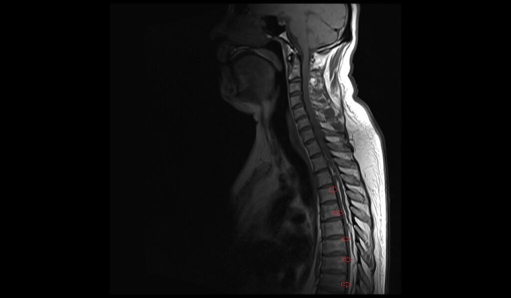
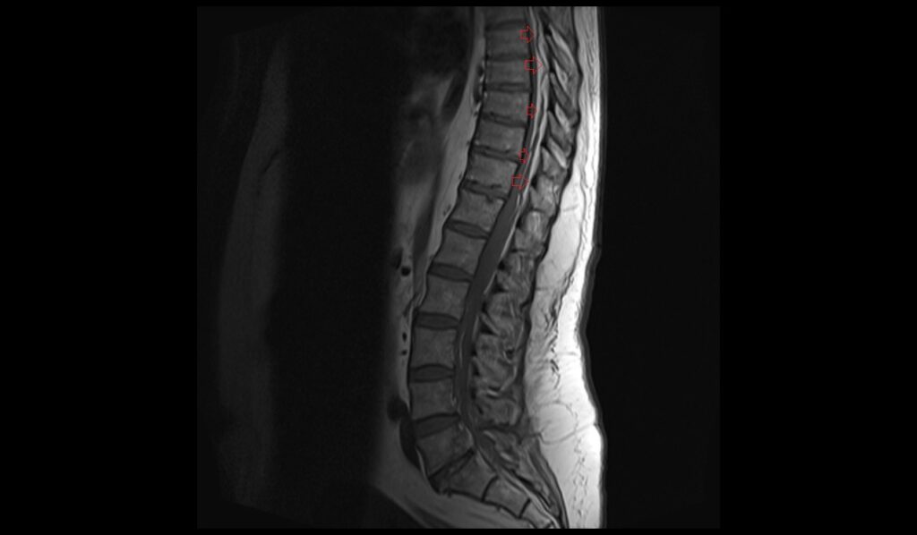
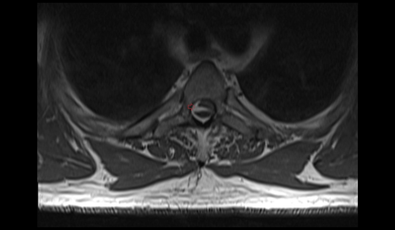
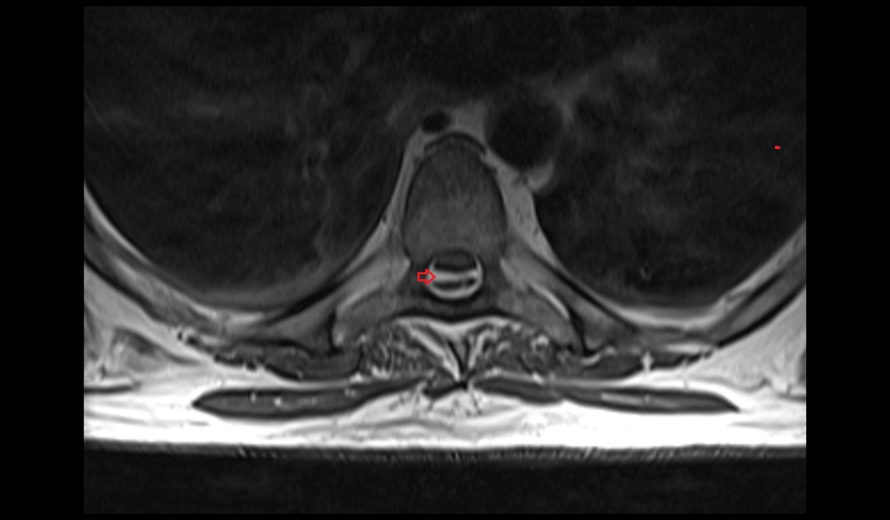
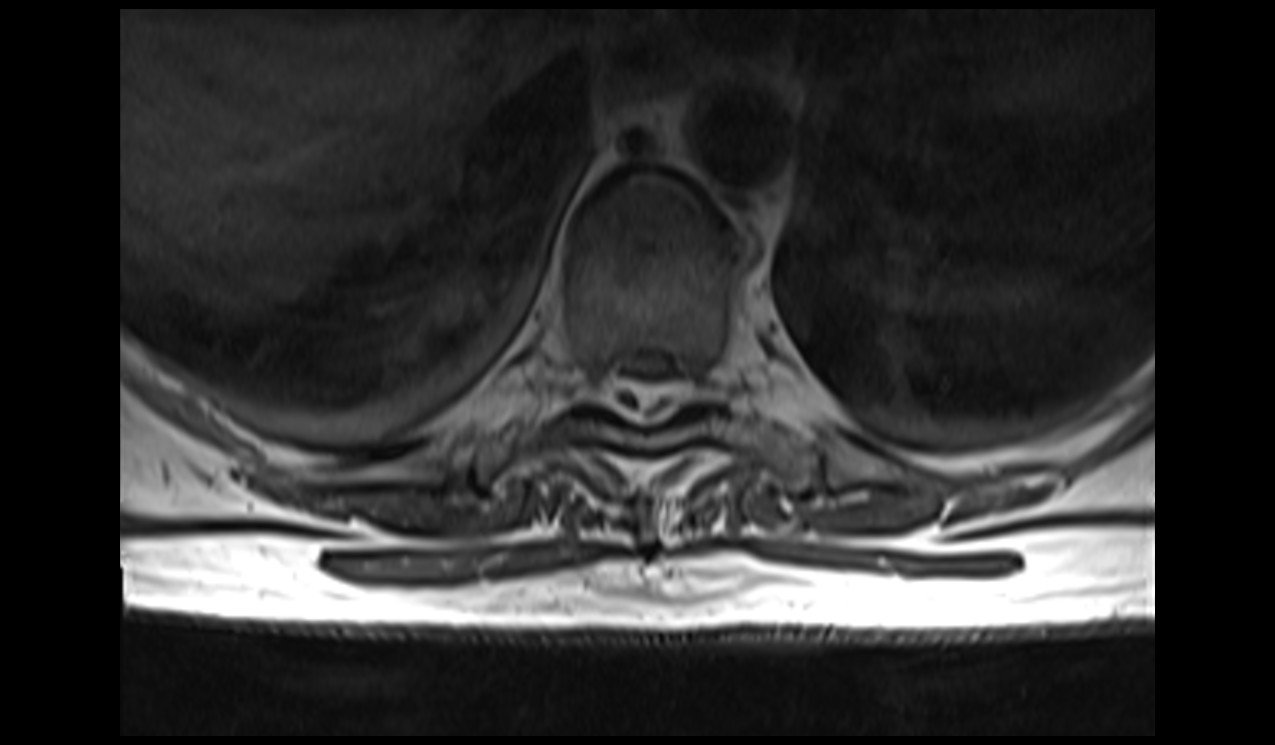
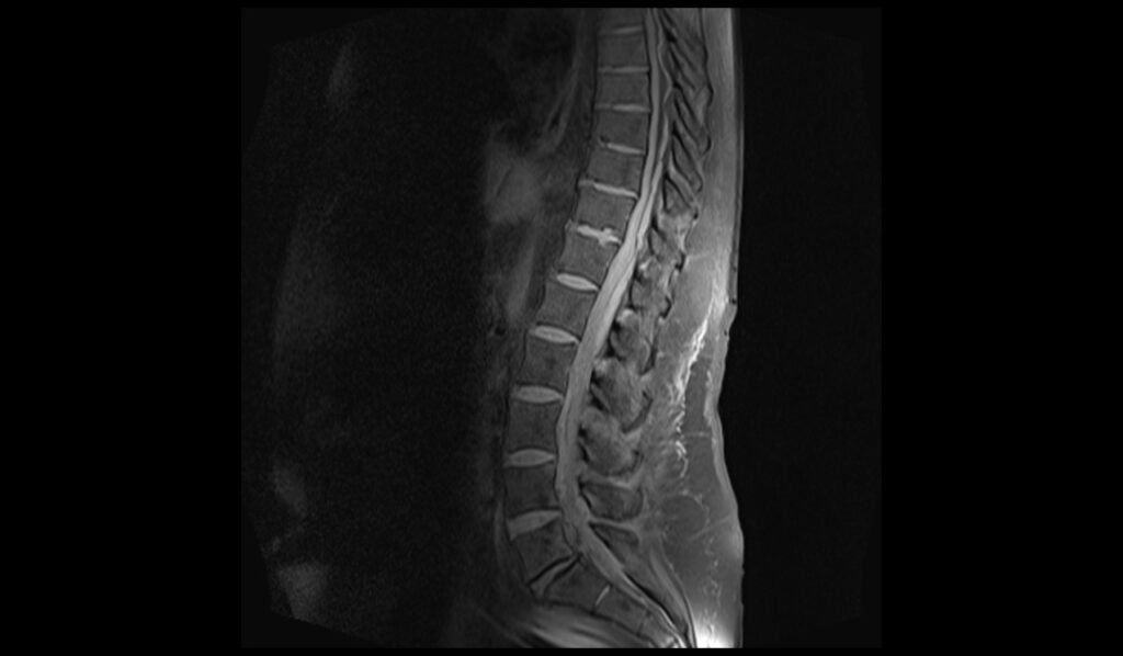
On T1-weighted MRI, a spinal epidural hematoma typically appears as a hyperintense (bright) or isointense (similar in intensity) lesion relative to the spinal cord. In the acute phase, the hematoma may appear isointense due to the presence of deoxyhemoglobin. As the hematoma evolves, the signal intensity increases due to the conversion of deoxyhemoglobin to methemoglobin, resulting in a hyperintense appearance. The precise location of the hematoma within the epidural space and its effect on adjacent structures, such as spinal cord compression, can be clearly visualized, aiding in the assessment of the severity and extent of the condition.
MRI T2 Appearance of Spinal Epidural Hematoma
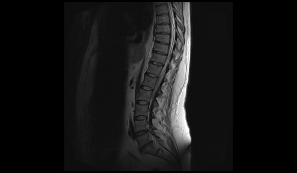
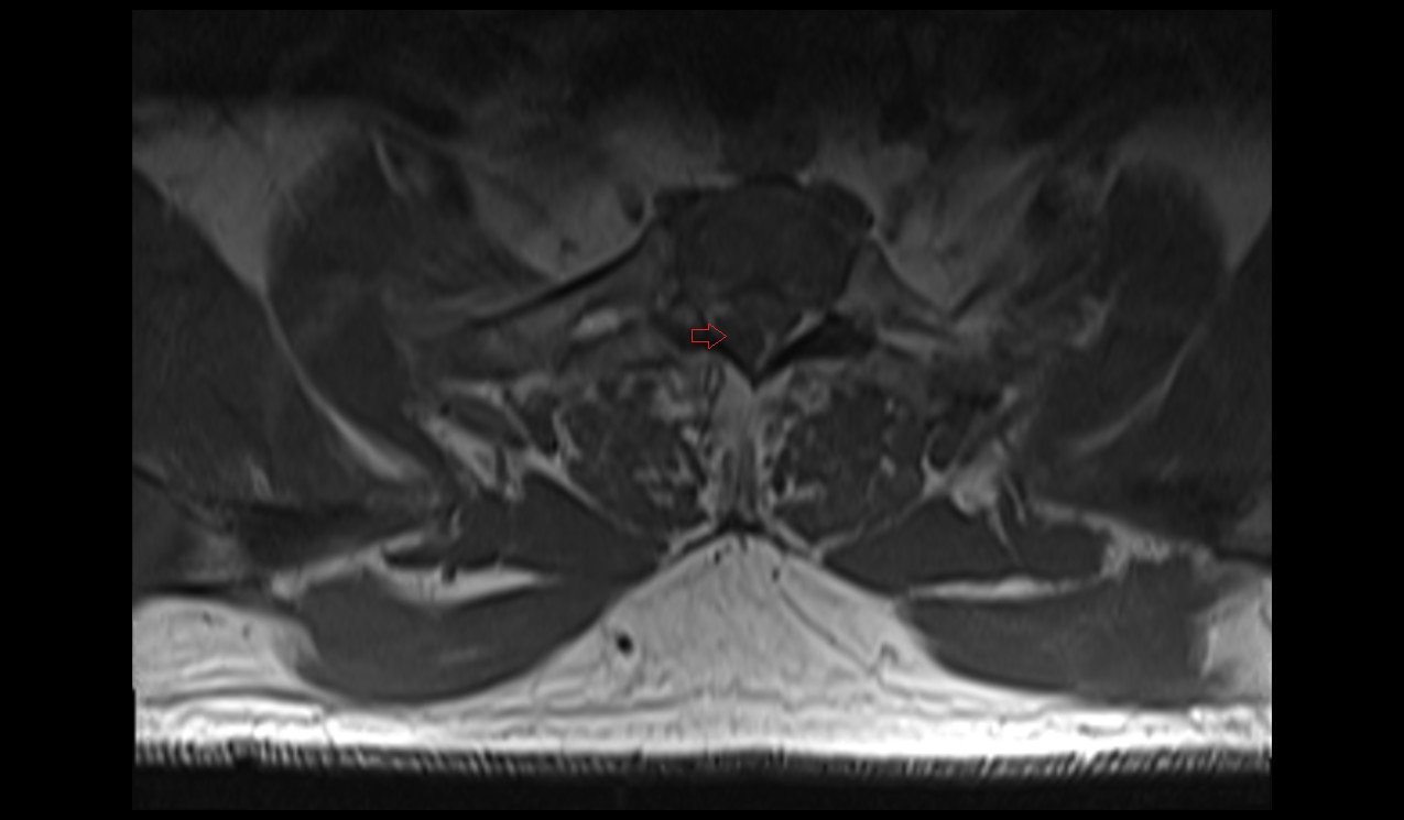
On T2-weighted MRI, a spinal epidural hematoma generally appears as a hyperintense (bright) lesion due to the high water content in the hematoma. This hyperintensity is often more pronounced in the subacute phase when the hematoma contains methemoglobin. In the acute phase, the hematoma may appear hypointense or isointense due to the presence of deoxyhemoglobin. T2-weighted images are particularly useful for identifying associated edema and inflammation in the surrounding tissues and spinal cord. The extent of the hematoma and its mass effect on the spinal cord and nerve roots can be well appreciated in this sequence.
MRI T1 Post-Contrast Appearance of Spinal Epidural Hematoma
On T1-weighted post-contrast MRI, a spinal epidural hematoma typically does not enhance because the hematoma itself is composed of blood products, which do not take up the contrast agent. However, there may be peripheral enhancement if there is associated inflammation or disruption of the epidural venous plexus. The surrounding dura mater may also enhance due to its vascular nature. Post-contrast imaging is useful for distinguishing the hematoma from other enhancing lesions or pathologies, such as abscesses or tumors, and for evaluating any potential complications or secondary effects related to the hematoma.
T1 sagittal image of cervical spine shows Spinal Epidural Hematoma
T1 sagittal image of lumbar spine shows Spinal Epidural Hematoma
T2 sagittal image of cervical spine shows Spinal Epidural Hematoma

T2 sagittal image of lumbar spine shows Spinal Epidural Hematoma
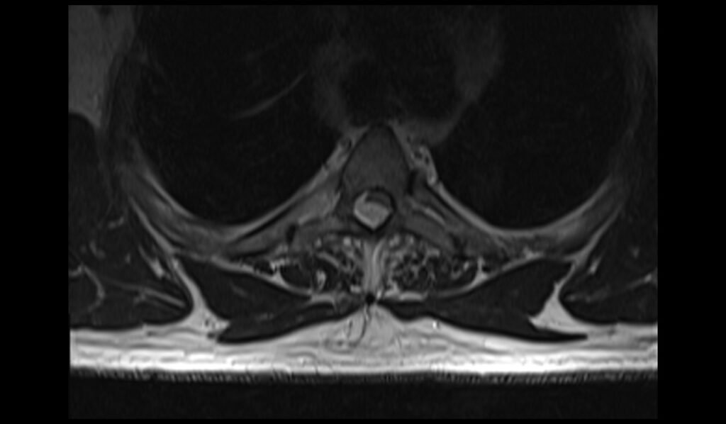
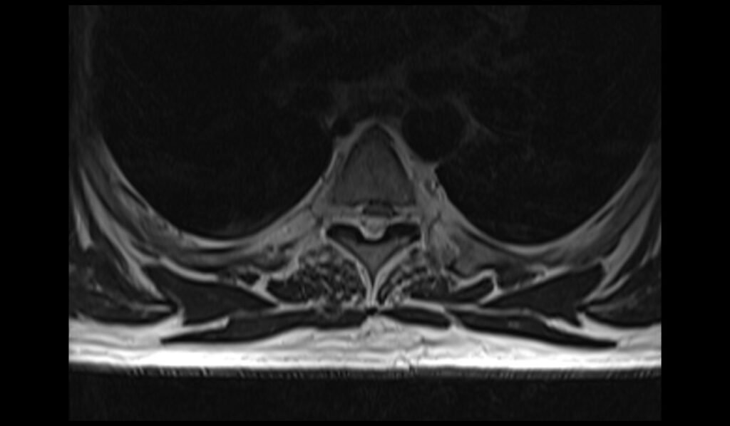
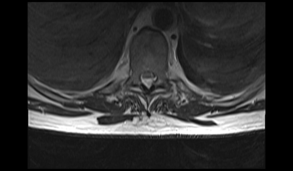
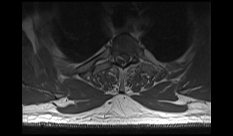
T2 TSE axial image of spine shows Epidural Hematoma

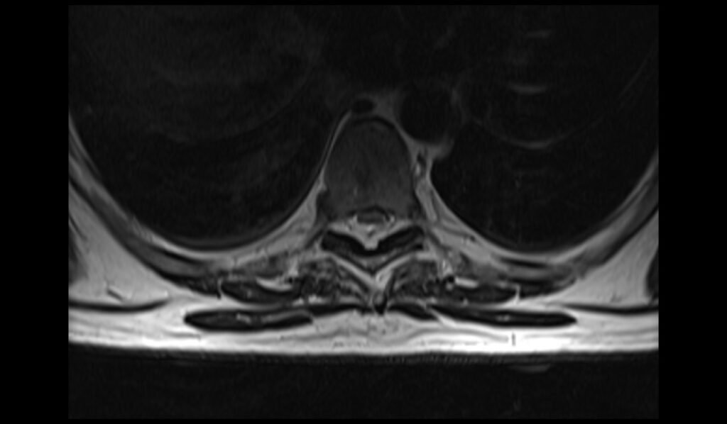
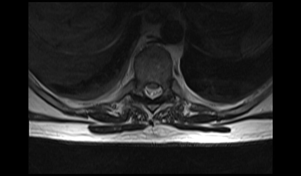
T1 TSE axial image of spine shows Epidural Hematoma

T1 FS post contrast sagittal image of cervical spine shows Spinal Epidural Hematoma

T1 FS post contrast sagittal image of lumbar spine shows Spinal Epidural Hematoma
References
- Alrazooqi, M. K., Skikic, E., Iqbal, S. S., Sulaiman, L., & Noori, O. Q. M. (2023). Traumatic spinal epidural hematoma with significant neurologic findings: A case report. Cureus, 15(5), e38869.
- Raasck, K., Habis, A. A., Aoude, A., Simões, L., Barros, F., Reindl, R., & Jarzem, P. (2017). Spontaneous spinal epidural hematoma management: A case series and literature review. Spinal Cord Series and Cases, 3, Article 16043.
- Moriarty, H. K., O Cearbhaill, R., Moriarty, P. D., Stanley, E., Lawler, L. P., & Kavanagh, E. C. (2019). MR imaging of spinal haematoma: A pictorial review. British Journal of Radiology, 92(1095), 20180532.
- Alrazooqi, M. K., Skikic, E., Iqbal, S. S., Sulaiman, L., & Muhammed Noori, O. Q. (2023). Traumatic spinal epidural hematoma with significant neurologic findings: A case report. Cureus, 15(5), e38869.
- Braun, P., Kazmi, K., Nogués-Meléndez, P., Mas-Estellés, F., & Aparici-Robles, F. (2007). MRI findings in spinal subdural and epidural hematomas. European Journal of Radiology, 64(1), 119-125.