MRI of Shoulder Metastasis
Shoulder metastasis refers to the spread of cancer cells from their original site to the shoulder region. This is a secondary cancer, meaning it originates elsewhere in the body and spreads to the shoulder.
Causes
The primary cause of shoulder metastasis is the spread of cancerous cells from a primary tumor site to the shoulder. This can occur through the bloodstream or lymphatic system. Common primary cancer sites that may metastasize to the shoulder include the breast, lung, prostate, kidney, and thyroid cancers.
Symptoms
- Pain: Persistent or worsening pain in the shoulder, which may increase at night or during activity.
- Swelling or Mass: Noticeable swelling or a palpable mass in the shoulder area.
- Decreased Mobility: Limited range of motion in the shoulder or arm.
- Neurological Symptoms: Tingling, numbness, or weakness in the arm or hand, if nerves are affected.
- Bone Fractures: Increased risk of fractures in the affected area due to weakened bone structure.
- General Cancer Symptoms: Weight loss, fatigue, and general malaise, especially if the metastasis is part of advanced cancer spread.
Treatment
The treatment of shoulder metastasis is often multifaceted and depends on the primary cancer type, the extent of the metastasis, the patient’s overall health, and other factors. Common treatment approaches include:
- Radiation Therapy: Used to shrink tumors and reduce pain.
- Surgery: In some cases, surgical intervention may be necessary to remove the tumor, relieve pressure on nerves, or stabilize the shoulder.
- Chemotherapy: Systemic treatment to target cancerous cells throughout the body.
- Targeted Therapy: Medications that specifically target cancer cells with certain characteristics.
- Physical Therapy: To maintain mobility and function in the shoulder and arm.
MRI Appearance of Shoulder Metastasis
T1-Weighted Images: On T1-weighted MRI, metastatic lesions often appear as areas of low signal intensity, which means they are darker compared to the normal high signal of fatty bone marrow. This contrast helps in identifying the lesions.
T2-Weighted Images: In T2-weighted images, metastatic lesions typically show high signal intensity, making them appear brighter than the surrounding bone. This is because the water content in these lesions is higher compared to normal bone tissue.
STIR (Short Tau Inversion Recovery): STIR is particularly sensitive to changes in water content. Metastatic lesions often appear hyperintense (very bright) on STIR images. This sequence is effective in suppressing fat signals, which enhances the visibility of lesions within the fatty bone marrow.
PD (Proton Density) Fat Saturation: In Proton Density Fat Sat images with fat saturation, the fat signal is suppressed, which helps to enhance the visibility of pathological lesions. Metastatic lesions often appear as areas of increased signal intensity on these images, contrasted against the dark background of suppressed fat.
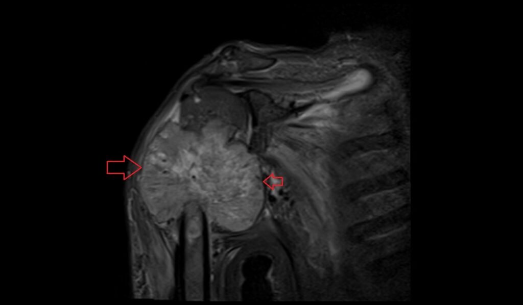
STIR coronal image shows shoulder metastasis
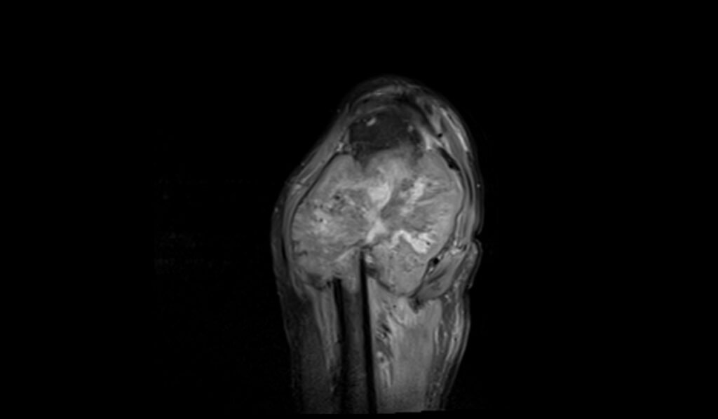
T1 TSE sagittal image shows shoulder metastasis

STIR axial image shows shoulder metastasis
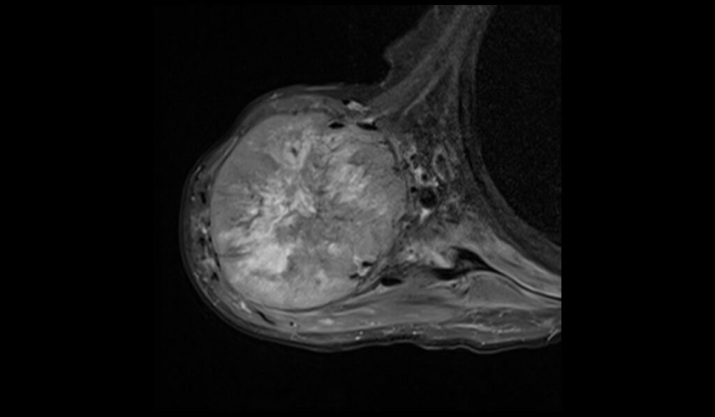
PD FS sagittal image shows shoulder metastasis
References
- Kim, S. Y., Jung, M. W., & Kim, J. M. (2011). The Shoulder Pain due to Metastatic Breast Cancer -A Case Report-. Korean Journal of Pain, 24(2), 119–122. https://doi.org/10.3344/kjp.2011.24.2.119
- O’Sullivan, G. J., Carty, F. L., & Cronin, C. G. (2015). Imaging of bone metastasis: An update. World Journal of Radiology, 7(8), 202–211. doi:10.4329/wjr.v7.i8.202
- Moore SL, Kransdorf MJ, Schweitzer ME, Murphey MD, Babb JS. “Can Sarcoidosis and Metastatic Bone Lesions Be Reliably Differentiated on Routine MRI?” AJR 2012; 198:1387–1393. American Roentgen Ray Society. DOI: 0361–803X/12/1986–1387.
- O’Sullivan, G. J., Carty, F. L., & Cronin, C. G. (2015). Imaging of bone metastasis: An update. World Journal of Radiology, 7(8), 202–211. https://doi.org/10.4329/wjr.v7.i8.202