Posterior Reversible Encephalopathy Syndrome (PRES) MRI
Posterior Reversible Encephalopathy Syndrome (PRES) is a neurological disorder characterized by a range of symptoms that arise due to the disruption of the blood-brain barrier, leading to vasogenic edema predominantly in the posterior regions of the brain. It is considered a reversible condition if promptly diagnosed and treated.
Causes
PRES can be triggered by a variety of factors, including:
- Hypertension: Severe or acute blood pressure elevations.
- Eclampsia/Pre-eclampsia: Conditions associated with pregnancy.
- Renal Failure: Chronic or acute kidney disease.
- Autoimmune Disorders: Such as systemic lupus erythematosus.
- Immunosuppressive Therapy: Medications like cyclosporine or tacrolimus.
- Infections and Sepsis: Severe systemic infections.
- Chemotherapy: Certain chemotherapeutic agents can precipitate PRES.
Symptoms
Symptoms of PRES can vary but often include:
- Headache: Severe, often sudden in onset.
- Seizures: Generalized or focal seizures.
- Visual Disturbances: Blurred vision, hemianopia, or cortical blindness.
- Altered Mental Status: Confusion, reduced consciousness, or coma.
- Nausea and Vomiting: Often associated with severe headaches.
- Weakness: Focal neurological deficits, sometimes mimicking a stroke.
Diagnosis
Diagnosis of PRES involves a combination of clinical evaluation and imaging studies:
- Clinical Assessment: Recognition of typical symptoms and potential triggering factors.
- Magnetic Resonance Imaging (MRI): The gold standard for diagnosis, showing characteristic findings of vasogenic edema in the parieto-occipital regions of the brain.
- Computed Tomography (CT): May show changes suggestive of PRES, though less sensitive than MRI.
- Blood Pressure Monitoring: Identifying severe hypertension.
- Laboratory Tests: To rule out other causes and identify potential triggers like renal failure or eclampsia.
Treatment
Management of PRES focuses on addressing the underlying cause and controlling symptoms:
- Blood Pressure Control: Aggressive management of hypertension with intravenous antihypertensive agents.
- Seizure Management: Antiepileptic drugs may be used to control seizures.
- Treatment of Underlying Conditions: Addressing eclampsia, renal failure, or cessation of offending medications.
- Supportive Care: Ensuring adequate hydration, monitoring neurological status, and supportive measures in intensive care if necessary.
MRI appearance of Posterior Reversible Encephalopathy Syndrome (PRES)
MRI T1 Appearance of Posterior Reversible Encephalopathy Syndrome (PRES)
On T1-weighted MRI images, Posterior Reversible Encephalopathy Syndrome (PRES) typically presents as hypointense (dark) regions within the affected areas of the brain. These hypointense areas are most commonly seen in the parieto-occipital regions, although the frontal and temporal lobes, cerebellum, and brainstem can also be involved. The T1 sequence is less sensitive for detecting the edema characteristic of PRES, but it can be useful for identifying any associated structural abnormalities or complications, such as hemorrhage, which may appear as hyperintense (bright) areas within the hypointense regions.
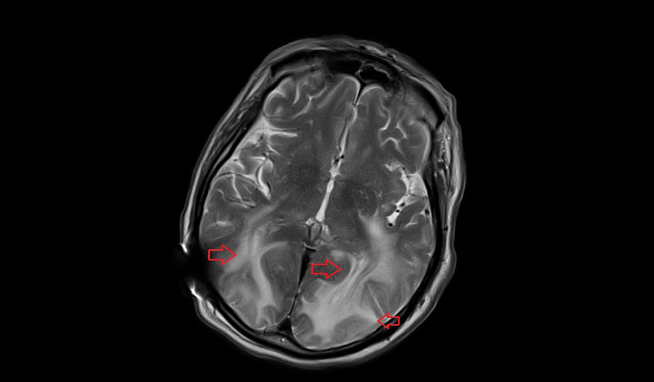
MRI T2 Appearance of Posterior Reversible Encephalopathy Syndrome (PRES)
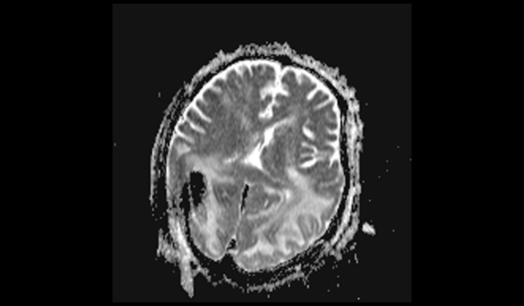
T2-weighted MRI images are highly sensitive for detecting the vasogenic edema associated with PRES. In patients with PRES, T2 images typically show hyperintense (bright) areas in the affected brain regions, most commonly in the parieto-occipital lobes. These hyperintense signals correspond to the accumulation of extracellular fluid, indicating the presence of edema. The T2 sequence effectively highlights the extent and severity of the edema, providing crucial information for diagnosis and monitoring the progression or resolution of the condition.
MRI FLAIR Appearance of Posterior Reversible Encephalopathy Syndrome (PRES)
Fluid-Attenuated Inversion Recovery (FLAIR) MRI sequences are particularly useful for evaluating PRES, as they suppress the cerebrospinal fluid (CSF) signal, enhancing the visibility of edema. In PRES, FLAIR images typically show hyperintense (bright) areas in the subcortical and cortical regions of the parieto-occipital lobes, as well as in other affected areas such as the frontal lobes, temporal lobes, cerebellum, and brainstem. These hyperintense areas indicate the presence of vasogenic edema and are more conspicuous on FLAIR images than on T2-weighted images, making FLAIR a critical sequence for diagnosing PRES.
MRI DWI b0, b1000, and ADC Appearance of Posterior Reversible Encephalopathy Syndrome (PRES)
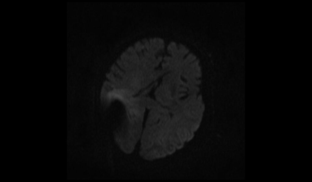
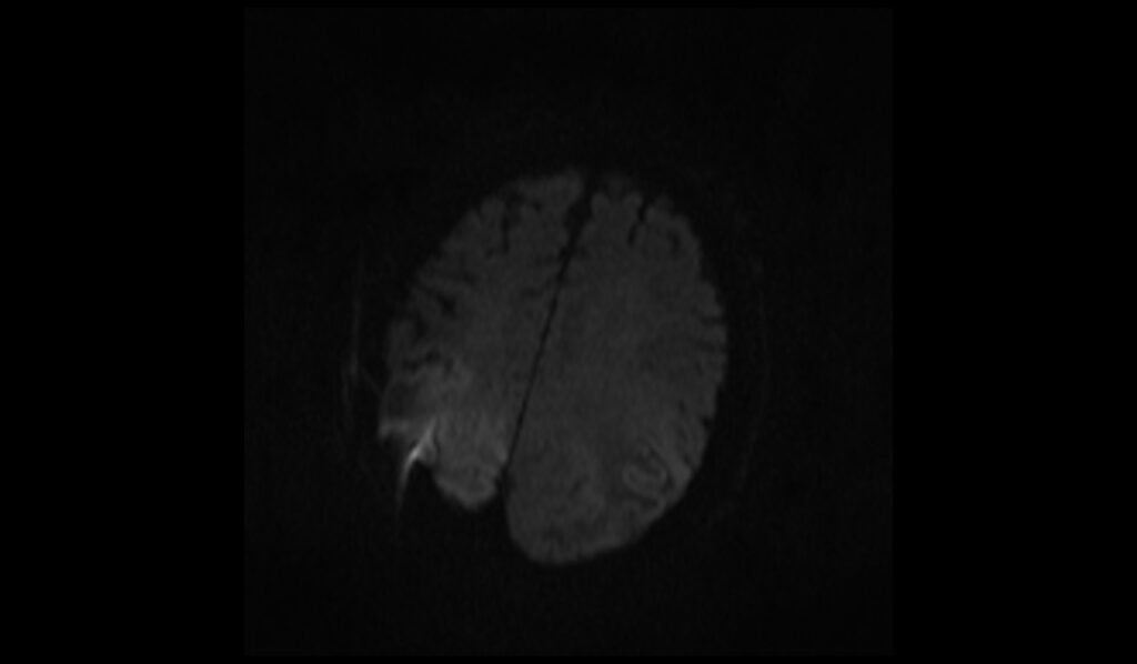
Diffusion-Weighted Imaging (DWI) and the Apparent Diffusion Coefficient (ADC) are important for differentiating between vasogenic and cytotoxic edema in PRES. On DWI with b0 and b1000 images, PRES typically shows isointense or mildly hyperintense signals, reflecting the presence of vasogenic edema rather than cytotoxic edema. The ADC maps show increased signal intensity in the affected areas, indicating increased diffusion of water molecules, which is characteristic of vasogenic edema. This helps distinguish PRES from conditions like acute ischemia, which would show restricted diffusion (hyperintensity on DWI and hypointensity on ADC).
MRI T1 Post-Contrast Appearance of Posterior Reversible Encephalopathy Syndrome (PRES)
On T1-weighted post-contrast MRI images, PRES may show variable enhancement patterns. In many cases, there is minimal to no enhancement, as vasogenic edema typically does not disrupt the blood-brain barrier significantly. However, in some instances, mild leptomeningeal or cortical enhancement may be observed, particularly if there is associated inflammation or breakdown of the blood-brain barrier. The post-contrast T1 sequence can help identify any complicating factors such as superimposed infection.
T2 axial images shows Posterior Reversible Encephalopathy Syndrome (PRES) of brain

FLAIR axial images shows Posterior Reversible Encephalopathy Syndrome (PRES) of brain

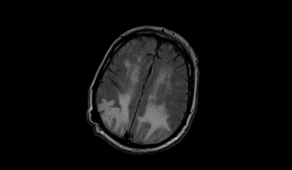
T1 axial images shows shows Posterior Reversible Encephalopathy Syndrome (PRES) of brain


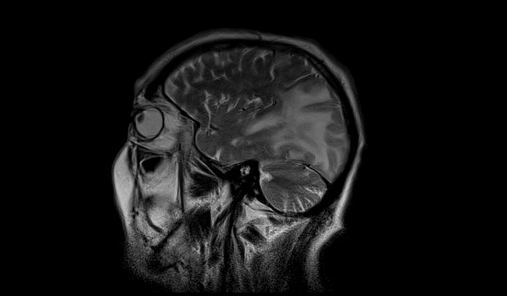
T2 sagittal image shows Posterior Reversible Encephalopathy Syndrome (PRES) of brain

DWI b0 image shows shows Posterior Reversible Encephalopathy Syndrome (PRES) of brain

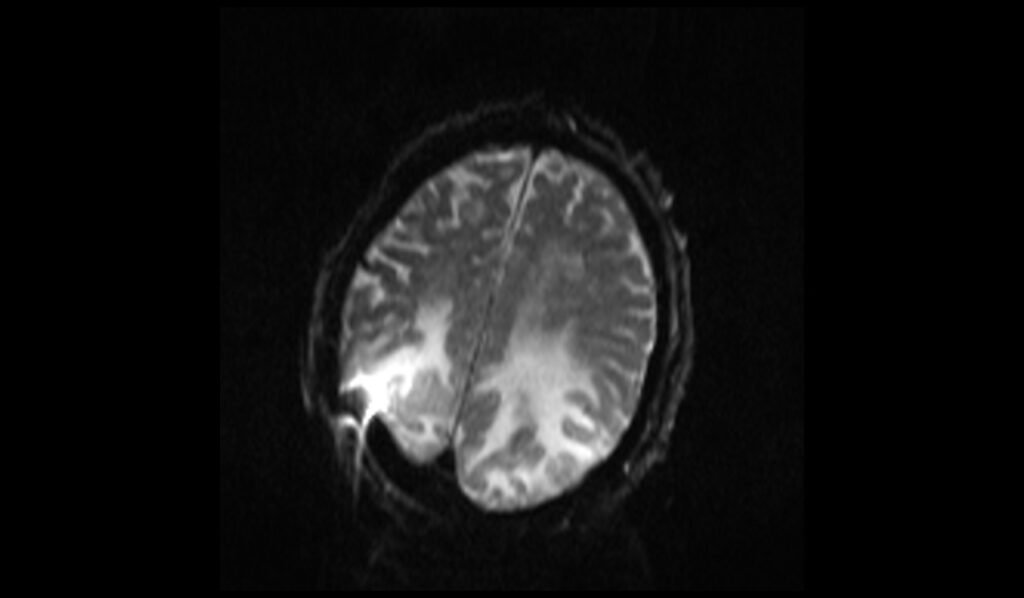
DWI b1000 image shows Posterior Reversible Encephalopathy Syndrome (PRES) of brain
DWI ADC map image shows Posterior Reversible Encephalopathy Syndrome (PRES) of brain

References
- Sudulagunta, S. R., Sodalagunta, M. B., Kumbhat, M., & Nataraju, A. S. (2017). Posterior reversible encephalopathy syndrome (PRES). Oxf Med Case Reports, 2017(4), omx011. https://doi.org/10.1093/omcr/omx011
- Hobson, E. V., Craven, I., & Blank, S. C. (2012). Posterior Reversible Encephalopathy Syndrome: A Truly Treatable Neurologic Illness. Peritoneal Dialysis International, 32(6), 590-594. doi: 10.3747/pdi.2012.00152. PMCID: PMC3524908. PMID: 23212858.
- Raman, R., Devaramane, R., Jagadish, G. M., & Chowdaiah, S. (2017). Various Imaging Manifestations of Posterior Reversible Encephalopathy Syndrome (PRES) on Magnetic Resonance Imaging (MRI). Polish Journal of Radiology, 82, 64–70. https://doi.org/10.12659/PJR.899960
- McKinney, A. M., Short, J., Truwit, C. L., McKinney, Z. J., Kozak, O. S., SantaCruz, K. S., & Teksam, M. (2007). Posterior reversible encephalopathy syndrome: Incidence of atypical regions of involvement and imaging findings. American Journal of Roentgenology, 189(4). https://doi.org/10.2214/AJR.07.2024
- Bartynski, W. S. (2008). Posterior reversible encephalopathy syndrome, part 1: Fundamental imaging and clinical features. American Journal of Neuroradiology, 29(6), 1036-1042.