MRI Pontine Glioma
Pontine Glioma, more specifically known as Diffuse Intrinsic Pontine Glioma (DIPG), is a type of brain tumor that occurs in the pons, which is a part of the brainstem. This condition primarily affects children, usually between the ages of 5 and 10, and is known for its aggressive nature and poor prognosis.
Causes
The exact cause of pontine gliomas, like many other cancers, remains largely unknown. However, research indicates that these tumors arise from glial cells, which are supportive cells in the brain.
Symptoms
Symptoms of pontine glioma typically develop quickly and may include:
- Problems with Eye Movement and Facial Expressions: Due to the tumor’s location in the brainstem, which controls many cranial nerves.
- Weakness in the Arms and Legs: This can sometimes be more pronounced on one side of the body.
- Difficulty in Walking and Coordination: Balance issues are common due to the impact on the brainstem.
- Changes in Speech: Difficulty in articulation or slurred speech.
- Headaches and Nausea: Caused by increased intracranial pressure.
Diagnosis
Diagnosis typically involves neurological examination and imaging studies, such as magnetic resonance imaging (MRI). The characteristic appearance of DIPG on MRI is often sufficient for diagnosis, and due to the tumor’s location, a biopsy is not always performed.
Treatment
Treating pontine gliomas is challenging due to their location and the tendency of the cancer cells to infiltrate surrounding brain tissue.
- Radiation Therapy: This is the primary treatment and can improve symptoms and quality of life, but it is not curative.
- Chemotherapy: Has limited effectiveness but may be used alongside radiation.
- Clinical Trials: Many patients participate in clinical trials that explore new treatment options, such as targeted therapies or immunotherapy.
MRI appearance of Pontine Glioma
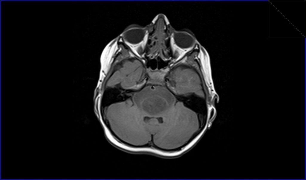
T1-Weighted Imaging (T1WI): Pontine gliomas typically appear hypointense or isointense on T1-weighted images. This means they are generally the same or a slightly darker shade compared to the normal brain tissue.
T2-Weighted Imaging (T2WI): On T2-weighted images, these tumors usually appear hyperintense, meaning they are brighter than the surrounding brain tissue. This is due to the high water content of the tumor.
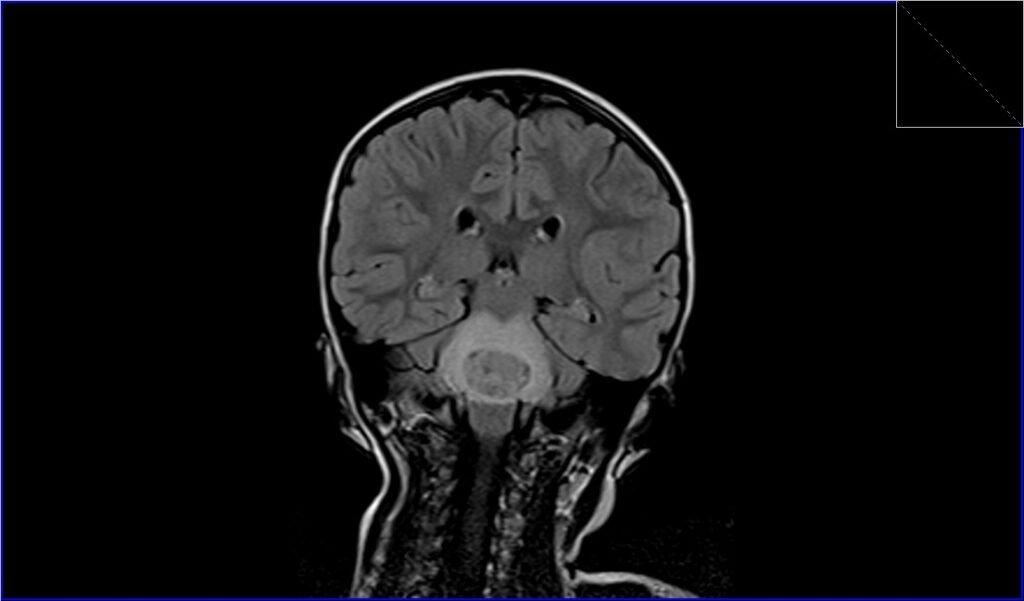
Fluid-Attenuated Inversion Recovery (FLAIR): In FLAIR imaging, pontine gliomas often appear hyperintense. The FLAIR sequence is particularly useful as it suppresses the signal from the cerebrospinal fluid, allowing better visualization of the tumor against the background of the brain.
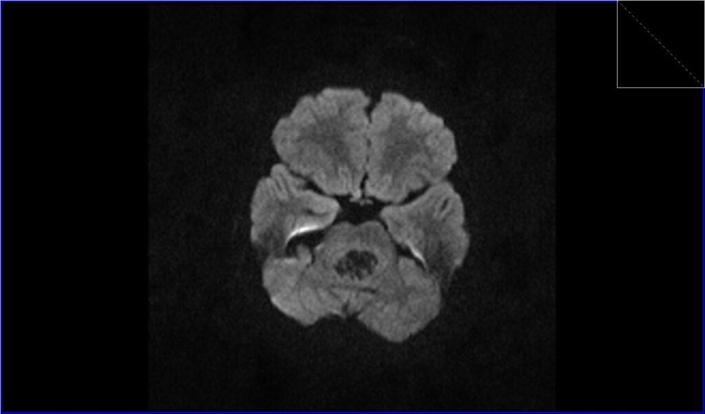
DWI (Diffusion Weighted Imaging): DWI assesses the movement of water molecules in tissue. Pontine gliomas can show varied intensity on DWI.
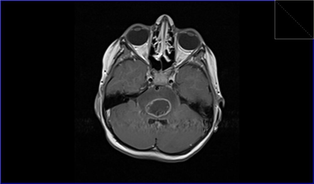
Post-Contrast T1-Weighted MRI: Pontine gliomas generally do not enhance uniformly. Some parts of the tumor may take up the contrast and appear brighter, while other parts may not, indicating a heterogeneous nature.
T2 axial image shows Pontine Glioma

FLAIR coronal image shows Pontine Glioma
T1 axial image shows Pontine Glioma
DWI b0 image shows Pontine Glioma

DWI b1000 image shows Pontine Glioma
DWI ADC map image shows Pontine Glioma

T1 post contrast axial image shows Medulloblastoma
References
- Mathew RK, Rutka JT. Diffuse Intrinsic Pontine Glioma: Clinical Features, Molecular Genetics, and Novel Targeted Therapeutics. J Korean Neurosurg Soc. 2018 May;61(3):343-351. doi: 10.3340/jkns.2018.0008. PMID: 29742880; PMCID: PMC5957322.
- Farrukh, S., Habib, S., Rafaqat, A., Sarfraz, Z., Sarfraz, A., Sarfraz, M., Robles-Velasco, K., Felix, M., Cherrez-Ojeda, I. (2023). Emerging Therapeutic Strategies for Diffuse Intrinsic Pontine Glioma: A Systematic Review. Healthcare (Basel), 11(4), 559.
- Tisnado, J., Young, R., Peck, K. K., & Haque, S. (2016). Conventional and Advanced Imaging of Diffuse Intrinsic Pontine Glioma. J Child Neurol, 31(12), 1386–1393. Published online 2016 Apr 12. doi: 10.1177/0883073816634855. PMID: 27071471. PMCID: PMC5659185. Available in PMC 2017 Oct 27.
- Hargrave, D., Chuang, N., & Bouffet, E. (2008). Conventional MRI cannot predict survival in childhood diffuse intrinsic pontine glioma. Journal of Neuro-Oncology, 86, 313-319.
- Warren, K. E. (2012). Diffuse intrinsic pontine glioma: poised for progress. Frontiers in Oncology, 2, 205. https://doi.org/10.3389/fonc.2012.00205