Pituitary Iron Overload (hemochromatosis) MRI
Pituitary iron overload refers to the accumulation of excess iron in the pituitary gland, a small gland at the base of the brain that plays a crucial role in regulating various hormones. This condition is often associated with systemic iron overload disorders, such as hemochromatosis or transfusion-dependent anemias.
Causes
Pituitary iron overload typically occurs due to:
- Hemochromatosis: A genetic disorder causing the body to absorb too much iron from the diet.
- Frequent Blood Transfusions: Conditions like thalassemia or sickle cell anemia that require regular blood transfusions can lead to excess iron accumulation.
- Iron Supplement Overuse: Excessive intake of iron supplements over a long period.
- Chronic Liver Diseases: Conditions such as chronic hepatitis or cirrhosis can also contribute to iron overload.
Symptoms
Symptoms of pituitary iron overload are primarily related to the impaired function of the pituitary gland and can include:
- Hypogonadism: Reduced function of the gonads, leading to low sex hormone levels, infertility, reduced libido, and menstrual irregularities in women.
- Growth Hormone Deficiency: Particularly in children, leading to growth retardation and short stature.
- Thyroid Dysfunction: Symptoms of hypothyroidism, such as fatigue, weight gain, and depression.
- Adrenal Insufficiency: Symptoms include fatigue, muscle weakness, weight loss, and low blood pressure.
Diagnosis
The diagnosis of pituitary iron overload involves:
- Medical History and Physical Examination: To identify risk factors and symptoms suggestive of iron overload.
- Blood Tests: To measure iron levels, including serum ferritin, transferrin saturation, and total iron-binding capacity.
- Hormone Levels: Assessing the levels of pituitary hormones like LH, FSH, TSH, ACTH, and growth hormone.
- Imaging Studies: Magnetic Resonance Imaging (MRI) is the most effective tool for detecting iron overload in the pituitary gland.
- Genetic Testing: To identify hereditary hemochromatosis.
Treatment
Treatment of pituitary iron overload aims to reduce iron levels and manage hormone deficiencies:
- Iron Chelation Therapy: Medications like deferoxamine, deferasirox, or deferiprone are used to bind excess iron and facilitate its excretion.
- Phlebotomy: Regular blood removal to reduce iron levels in patients with hereditary hemochromatosis.
- Hormone Replacement Therapy: To address deficiencies in sex hormones, thyroid hormones, or adrenal hormones.
- Lifestyle Modifications: Diet low in iron and avoidance of iron supplements and vitamin C (which increases iron absorption).
MRI appearance of Pituitary iron overload
T1-Weighted MRI appearance of Pituitary iron overload
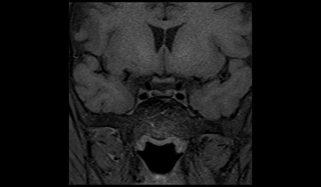
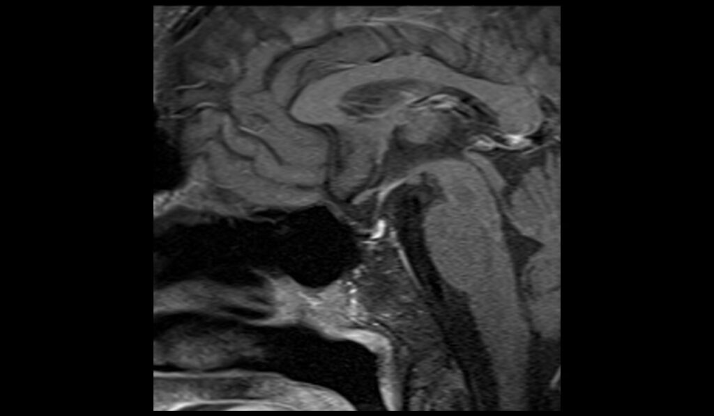
In patients with pituitary iron overload, T1-weighted MRI sequences typically show low signal intensity in the pituitary gland. Iron deposits within the pituitary result in a shortened T1 relaxation time, leading to a darker appearance of the gland on T1 images. This characteristic hypointensity helps differentiate areas of iron accumulation from the surrounding normal tissue, which appears relatively brighter.
T2-Weighted MRI appearance of Pituitary iron overload
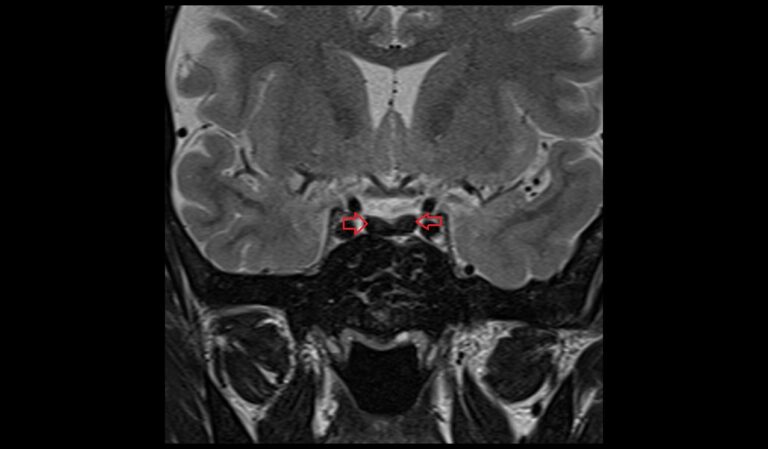
T2-weighted MRI sequences also exhibit low signal intensity in cases of pituitary iron overload. Similar to T1, the presence of iron causes a reduction in T2 relaxation time, making the pituitary gland appear darker compared to other brain structures. This effect is more pronounced on T2 than on T1, providing a clearer contrast and making T2-weighted imaging particularly useful for assessing the extent of iron deposition.
T1 Post-Contrast MRI appearance of Pituitary iron overload
Following the administration of contrast material, T1 post-contrast images of a pituitary with iron overload generally do not show significant enhancement. The presence of iron can hinder the uptake of contrast, resulting in minimal or no change in the signal intensity of the gland post-contrast. This lack of enhancement can be crucial for diagnosing iron overload, as it contrasts with other pituitary abnormalities, such as adenomas or inflammatory conditions, which typically show enhancement after contrast administration.
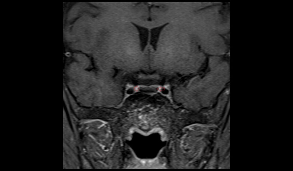
T2 coronal images shows Pituitary Iron Overload
T1 coronal images shows Pituitary Iron Overload
T1 sagittal images shows Pituitary Iron Overload
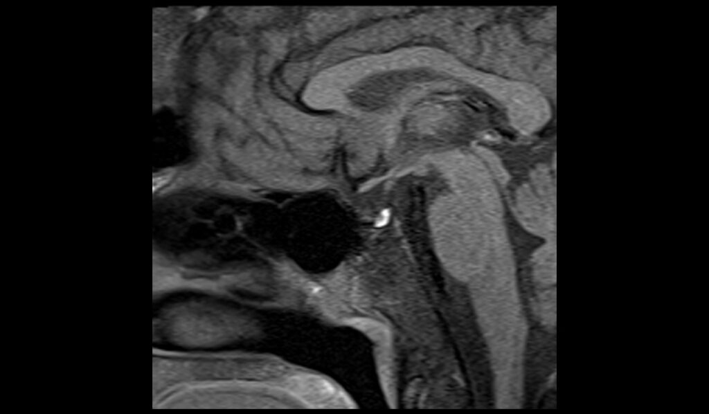
T1 post contrast sagittal images shows Pituitary Iron Overload
T1 post contrast coronal images shows Pituitary Iron Overload
References
- Tessi, R. T. Y., Oze, R. K., Outznit, M., Hsain, I. H., Nassar, I., & Billah, N. M. (2023). Pituitary hyposignal characteristic of hemochromatosis on MRI. Clinical Case Reports, 11(1), e6384. https://doi.org/10.1002/ccr3.6384
- Argyropoulou, M. I., Metafratzi, Z., Kiortsis, D. N., Bitsis, S., Tsatsoulis, A., & Efremidis, S. (2000). T2 relaxation rate as an index of pituitary iron overload in patients with β-thalassemia major. American Journal of Roentgenology, 175(6). https://doi.org/10.2214/ajr.175.6.1751567
- Verberckmoes, B., Dekeyzer, S., & Decaestecker, K. (2024). The dark pituitary: Hemochromatosis as a lesser-known cause of pituitary dysfunction. Journal of the Belgian Society of Radiology. Retrieved from Journal of the Belgian Society of Radiology website.