Perianal fistula MRI
A perianal fistula is an abnormal, tube-like connection that forms between the inside of the anus and the skin near the anus. It typically results from an infection in an anal gland that spreads to the skin, causing an abscess.
Causes
The primary cause of a perianal fistula is an infection in the anal gland. Other potential causes include:
- Crohn’s Disease: A chronic inflammatory bowel disease that can cause fistulas in the gastrointestinal tract.
- Ulcerative Colitis: Another form of inflammatory bowel disease.
- Trauma: Injury to the anal region.
- Infections: Such as tuberculosis or sexually transmitted infections.
Symptoms
The symptoms of a perianal fistula can vary, but common ones include:
- Pain: Around the anus, which can be severe.
- Swelling: Around the anus.
- Discharge: Of pus or blood from an opening near the anus.
- Irritation: Of the skin around the anus.
- Recurrent Anal Abscesses: Chronic or recurrent abscesses in the perianal region.
- Difficulty Sitting: Due to pain and swelling.
Diagnosis
Diagnosis typically involves:
- Physical Examination: By a doctor to inspect the area around the anus.
- Anoscopy: A procedure where a small, tube-like instrument is inserted into the anus to view the anal canal.
- Imaging Tests:
- MRI: To get detailed images of the fistula and surrounding structures.
- Fistulography: An X-ray procedure where a contrast dye is injected into the fistula to map its course.
Treatment
Treatment options vary depending on the complexity and severity of the fistula but can include:
- Surgery:
- Fistulotomy: The most common procedure, where the fistula tract is cut open and allowed to heal.
- Seton Placement: A surgical thread is placed in the fistula to keep it open and allow it to drain, reducing infection and promoting healing.
- Advancement Flap Procedure: Involves covering the internal opening of the fistula with a flap of tissue.
- Medications: Antibiotics to treat infection, and anti-inflammatory drugs if the fistula is associated with conditions like Crohn’s disease.
- Fibrin Glue: Injecting a glue into the fistula tract to close it
MRI Appearance of Perianal fistula
T2 Appearance of Perianal Fistula
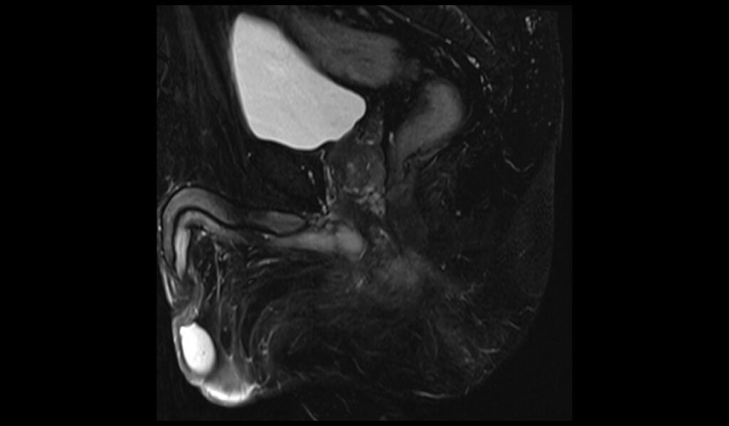
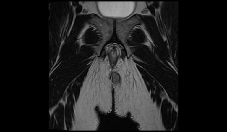
On T2-weighted MRI, perianal fistulas typically appear as hyperintense (bright) linear or branching structures due to their fluid content. The high water content within the fistula tract and any associated abscesses result in the increased signal intensity. Additionally, the surrounding inflamed tissue may also appear hyperintense, aiding in the delineation of the fistulous path. T2-weighted images are particularly useful for identifying the extent and complexity of the fistula, including secondary tracts and potential abscess formations.
STIR Appearance of Perianal Fistula
STIR (Short Tau Inversion Recovery) sequences are highly sensitive to fluid and inflammation, making them particularly useful for evaluating perianal fistulas. On STIR images, perianal fistulas typically exhibit high signal intensity due to the presence of fluid and inflamed tissues within and around the fistula tract. This sequence helps in suppressing fat signal, thereby enhancing the visibility of the fistula against the background of fatty tissues. The increased contrast between the fistula and surrounding structures allows for a clearer and more detailed assessment of the inflammatory process and any associated abscesses.
T1 Appearance of Perianal Fistula
On T1-weighted MRI, perianal fistulas generally appear as hypointense (dark) structures due to the low signal intensity of the fluid within the fistula tract. In contrast, the surrounding tissues, particularly fat, appear hyperintense (bright), providing a clear differentiation from the fistulous path. T1-weighted images are less sensitive to fluid and inflammation compared to T2 or STIR sequences, but they are useful in identifying the anatomical relationships and the extent of the fistula in the context of the surrounding pelvic structures. Additionally, post-contrast T1-weighted images can enhance the visualization of active inflammation and help in detecting any abscesses by highlighting areas of contrast enhancement.
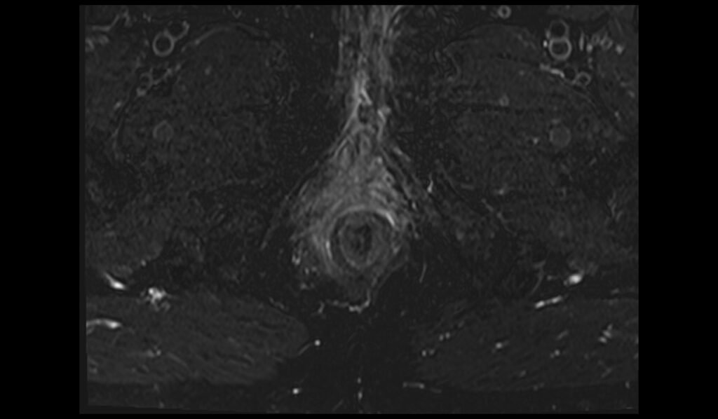
STIR axial oblique image of pelvis shows Perianal Fistula.


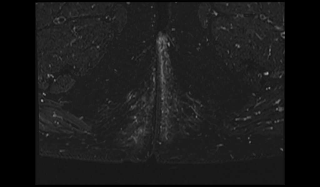
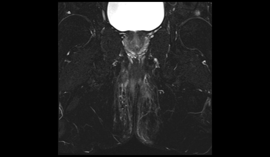
STIR coronal oblique image of pelvis shows Perianal Fistula.


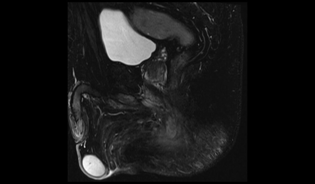
STIR sagittal oblique image of pelvis shows Perianal Fistula.


T2 axial oblique image of pelvis shows Perianal Fistula.


T2 coronal oblique image of pelvis shows Perianal Fistula.


Case Study 2
T2 FS Axial oblique

T2 FS coronal oblique

T2 FS sagittal

Case Study 3
T2 FS Axial oblique


T2 FS coronal oblique

T2 FS sagittal


References
Torkzad, M. R., & Karlbom, U. (2010). MRI for assessment of anal fistula. Insights into Imaging, 1(2), 62-71. doi:10.1007/s13244-010-0022-y. PMCID: PMC3259332. PMID: 22347906.- Gage, K. L., Deshmukh, S., Macura, K. J., Kamel, I. R., & Zaheer, A. (2013). MRI of perianal fistulas: Bridging the radiologic-surgical divide. Abdominal Imaging, 38(5), 1033-1042.
- Criado, J. de M., García del Salto, L., Fraga Rivas, P., Aguilera del Hoyo, L. F., Gutiérrez Velasco, L., Díez Pérez de las Vacas, M. I., Marco Sanz, A. G., Manzano Paradela, M., & Fraile Moreno, E. (2011). MR imaging evaluation of perianal fistulas: Spectrum of imaging features. RadioGraphics, 32(1). https://doi.org/10.1148/rg.321115040
- Joshi, A. R., & Siledar, S. G. (2014). Role of MRI in ano-rectal fistulas. Current Radiology Reports, 2, 63. https://doi.org/10.1007/s40134-014-0063-5