MRI Meningiomas
Meningiomas are tumors that originate from the meninges, which are the layers of tissue covering the brain and spinal cord. These tumors are typically slow-growing and are usually benign (non-cancerous) in nature. However, some meningiomas can exhibit more aggressive behavior and rare cases can be malignant (cancerous).
Meningiomas are the most common type of primary brain tumor in adults. They are often classified based on their appearance under a microscope and can have various subtypes. These tumors can arise from different locations along the meninges and can press against the brain or spinal cord as they grow, potentially causing neurological symptoms depending on their size and location.
Common symptoms of meningiomas include headaches, seizures, changes in vision, hearing loss, balance problems, weakness, and numbness in the limbs, among others. The severity of symptoms depends on the tumor’s location and size.
Meningiomas are typically diagnosed through imaging techniques such as MRI and CT scans, which can show the location, size, and characteristics of the tumor. Treatment options include observation (for slow-growing and asymptomatic tumors), surgical removal, radiation therapy, and in some cases, targeted drug therapy.
MRI appearance of Meningiomas
The appearance of a meningioma on MRI can vary depending on factors such as the tumor’s location, size, subtype, and other characteristics. Here are the key observed features in the MRI of meningiomas:
- T1-Weighted Images: Meningiomas typically appear iso- to hypointense on T1-weighted images. This means they often have similar or slightly lower signal intensity compared to the adjacent brain tissue. The exact appearance can vary based on factors like tumor composition and the presence of calcifications.
- T2-Weighted Images: On T2-weighted images, meningiomas often appear hyperintense. This hyperintensity is due to the relatively high water content in these tumors. The tumor’s appearance can sometimes be heterogeneous, with areas of varying signal intensity.
- FLAIR Images: In Fluid-Attenuated Inversion Recovery (FLAIR) images, meningiomas generally present as hyperintense lesions, emphasizing their relatively high water content.
- DWI Images: In Diffusion-Weighted Imaging (DWI), meningiomas typically appear hyperintense, indicating restricted diffusion due to their cellular structure. This heightened signal contrasts with the surrounding brain tissue.
- Post-Contrast Images (Gadolinium-Enhanced Images): One of the distinctive features of meningiomas is their avid uptake of contrast material (gadolinium) on MRI. After the administration of contrast, meningiomas typically enhance strongly and uniformly. The degree of enhancement can provide information about the tumor’s vascularity and characteristics.
- Tumor Margins: Meningiomas often have well-defined margins, which can help differentiate them from other types of brain tumors. This clear demarcation between the tumor and surrounding brain tissue is one of the features that make them distinguishable on MRI.
- Dural Tail: A characteristic feature associated with many meningiomas is the “dural tail.” This is an area of enhancement extending from the tumor into the adjacent dura mater (the outermost layer of the meninges). The dural tail is thought to be caused by reactive changes in the adjacent dura due to the tumor.

T2 Axial

Flair Axial

T1 Coronal Pre Contrast

AXI DWI B0
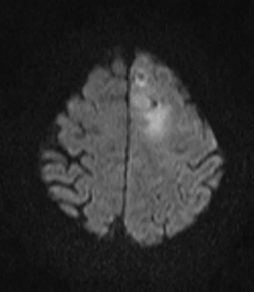
AXI DWI B 1000

AXI DWI ADC

T1 Coronal Post Contrast

T1 Axial Post Contrast
Case study 2


Case study 3


References
Watts, J., Box, G., Galvin, A., Brotchie, P., Trost, N., & Sutherland, T. (2014). Magnetic resonance imaging of meningiomas: A pictorial review. Insights into Imaging, 5(1), 113-122.
- Krishnan, V., Mittal, M. K., Sinha, M., & Thukral, B. B. (2019). Imaging spectrum of meningiomas: a review of uncommon imaging appearances and their histopathological and prognostic significance. Polish Journal of Radiology, 84, e630–e653.
- LaBella, D., Khanna, O., McBurney-Lin, S., Mclean, R., Nedelec, P., Rashid, A. S., Tahon, N. H., Altes, T., Baid, U., Bhalerao, R., Dhemesh, Y., Floyd, S., Godfrey, D., Hilal, F., Janas, A., Kazerooni, A., Kent, C., Kirkpatrick, J., Kofler, F., Leu, K., Maleki, N., Menze, B., Pajot, M., Reitman, Z. J., & Calabrese, E. (2024). A multi-institutional meningioma MRI dataset for automated multi-sequence image segmentation. Scientific Data, 11, Article 496. https://doi.org/10.1038/s41597-024-01987-0
- Ogasawara, C., Philbrick, B. D., & Adamson, D. C. (2021). Meningioma: A review of epidemiology, pathology, diagnosis, treatment, and future directions. Biomedicines, 9(3), 319.
- Hanna Jr, C., Willman, M., Cole, D., Mehkri, Y., Liu, S., Willman, J., & Lucke-Wold, B. (2023). Review of meningioma diagnosis and management. Egyptian Journal of Neurosurgery, 38(16).