Acute MCA Infarction
Stroke is described as a sudden neurological deficit that results from a cerebrovascular event. The middle cerebral artery (MCA) is the largest cerebral artery and is most commonly affected by a cerebrovascular accident. A middle cerebral artery (MCA) stroke is the sudden onset of a focal neurologic deficit caused by infarction or ischemia in the territory supplied by the middle cerebral artery. MRI with diffusion-weighted imaging is highly sensitive for the early diagnosis of MCA infarction.
After 24 hours post-stroke, middle cerebral artery (MCA) strokes appear as low signal intensity on T1-weighted images and as high signal intensity on T2-weighted and FLAIR images. Infarctions are hyperintense on T2 and FLAIR images due to the development of cytotoxic and vasogenic edema in the stroke area after 24 hours.
MRI Appearance of Middle Cerebral Artery (MCA) Stroke
Diffusion-Weighted Imaging (DWI): DWI is highly sensitive to changes in water diffusion caused by cellular edema. In acute MCA infarction, affected brain tissue exhibits hyperintensity on DWI due to restricted diffusion. This is a hallmark of the early stages of infarction and can be detected within minutes to hours after the onset of symptoms.
Apparent Diffusion Coefficient (ADC) Maps: ADC maps are derived from DWI data and provide quantitative information about the diffusion properties of tissues. In MCA infarctions, ADC values are decreased within the affected region, reflecting reduced diffusion caused by cellular swelling.
T2-Weighted Imaging: T2-weighted imaging can reveal areas of edema surrounding the infarcted tissue. The infarcted core appears hyperintense, while the surrounding edema appears hypointense.
Fluid-Attenuated Inversion Recovery (FLAIR): FLAIR sequences suppress the signal from cerebrospinal fluid, enhancing the visibility of perilesional edema and making the infarcted area more distinct. Infarcted regions appear hyperintense on FLAIR images.
- T1-Weighted Imaging: The infarcted region usually appears hypointense compared to the surrounding normal brain tissue. This hypointensity is due to changes in tissue composition, including cellular damage and edema.
- T1-Weighted Imaging with Gadolinium Contrast: Gadolinium-based contrast agents are often used to assess the perfusion status of brain tissue. In acute MCA infarction, there might be a delay in contrast arrival to the infarcted area, leading to a perfusion deficit.
Typical appearance of affected area in the event of early stroke
T2 and FLAIR images will be normal
T1 images will be normal
DWI b value 0 will be normal
DWI b value 1000 will be hyperintense
ADC map will be hypointense
Typical appearance of affected area 24 hours post stroke
T2 and FLAIR images will be hyperintense
T1 images will be hypointense
DWI b value 0 will be hyperintense
DWI b value 1000 will be hyperintense
ADC map will be hypointense

T2 AXIAL
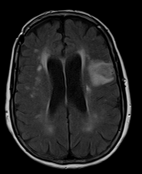
FLAIR AXIAL
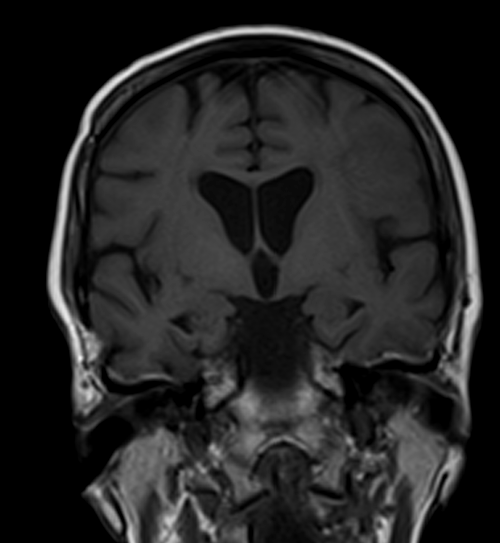
T1 CORONAL PRE CONTRAST

AXI DWI B 1000

AXI DWI ADC
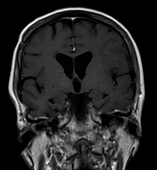
T1 CORONAL POST CONTRAST
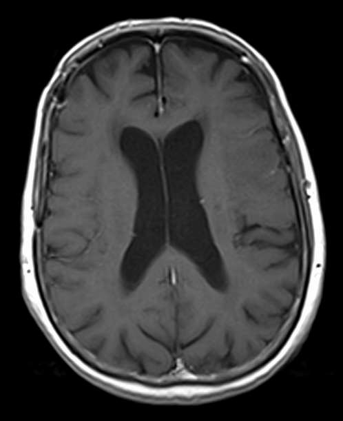
T1 AXIAL POST CONTRAST
References
Wardlaw, J. M., & Sellar, R. J. (2018). A simple practical classification of cerebral infarcts on MRI: PRODIGI. Radiography, 24(3), 238-245.
Schaefer, P. W. (2013). Acute ischemic stroke imaging: a practical approach for diagnosis and treatment decision-making. Neurologic Clinics, 31(3), 503-533.
Wintermark, M., Albers, G. W., Broderick, J. P., Demchuk, A. M., Fiebach, J. B., Fiehler, J., … & Warach, S. (2008). Acute Stroke Imaging Research Roadmap II. Stroke, 39(5), 1621-1628.
Thomalla, G., Cheng, B., Ebinger, M., Hao, Q., Tourdias, T., Wu, O., … & Gerloff, C. (2011). DWI-FLAIR mismatch for the identification of patients with acute ischaemic stroke within 4· 5 h of symptom onset (PRE-FLAIR): a multicentre observational study. The Lancet Neurology, 10(11), 978-986.
Kidwell, C. S., Chalela, J. A., Saver, J. L., Starkman, S., Hill, M. D., Demchuk, A. M., … & Warach, S. (2004). Comparison of MRI and CT for detection of acute intracerebral hemorrhage. Jama, 292(15), 1823-1830.
Nael, K., Trouard, T. P., Lafleur, S. R., Krupinski, E. A., Salamon, N., Kidwell, C. S., … & Villablanca, J. P. (2015). White matter ischemia in the vicinity of glioblastoma multiforme: correlation of DTI and PWI. Journal of Neuroimaging, 25(4), 628-635.