MRI of Nitrous Oxide Toxicity in the Spinal Cord
Nitrous Oxide Toxicity occurs when there is excessive exposure to or inhalation of nitrous oxide (N2O), commonly known as “laughing gas.” While N2O is widely used in medical settings for its anesthetic and analgesic properties, recreational use or occupational exposure can lead to toxicity.
Causes
- Medical Use: Overexposure during anesthesia or improper administration.
- Recreational Use: Inhaling nitrous oxide for its euphoric effects.
- Occupational Exposure: Inadequate ventilation in environments where nitrous oxide is used.
Symptoms
- Neurological: Tingling or numbness in extremities, weakness, ataxia (lack of muscle control), cognitive impairment, and in severe cases, myeloneuropathy (spinal cord and peripheral nerve damage).
- Psychological: Confusion, mood swings, hallucinations, and in severe cases, psychosis.
- Hematological: Megaloblastic anemia (enlarged red blood cells), due to interference with vitamin B12.
- Gastrointestinal: Nausea, vomiting, and abdominal pain.
Diagnosis
- Clinical History: Assessing exposure to nitrous oxide, either occupationally or recreationally.
- Neurological Examination: Checking for signs of neuropathy or myelopathy.
- Blood Tests: Measuring vitamin B12 levels, methylmalonic acid (MMA), and homocysteine levels. Elevated MMA and homocysteine are indicative of B12 deficiency.
- MRI: Imaging of the brain and spinal cord may be performed to identify any neurological damage.
Treatment
- Cessation of Exposure: Immediate discontinuation of nitrous oxide exposure.
- Vitamin B12 Supplementation: Intramuscular injections of vitamin B12 are often required, especially in cases of significant deficiency.
- Supportive Care: Symptomatic treatment for neurological and psychological symptoms.
- Monitoring: Regular follow-up to assess recovery of neurological function and normalization of blood parameters.
MRI Appearance of Nitrous Oxide Toxicity in the Spinal Cord
MRI T1 Appearance of Nitrous Oxide Toxicity in the Spinal Cord
On T1-weighted MRI images, nitrous oxide toxicity in the spinal cord may manifest as areas of hypointensity (darker signals) within the affected regions. These hypointense areas typically correspond to myelin damage or necrosis caused by the toxic effects of nitrous oxide on the nervous tissue. Additionally, T1-weighted images may reveal atrophy or structural changes in the spinal cord if the toxicity has led to significant neuronal loss or degeneration over time. T1 imaging is crucial for assessing the anatomical integrity and overall structural changes in the spinal cord due to nitrous oxide toxicity.
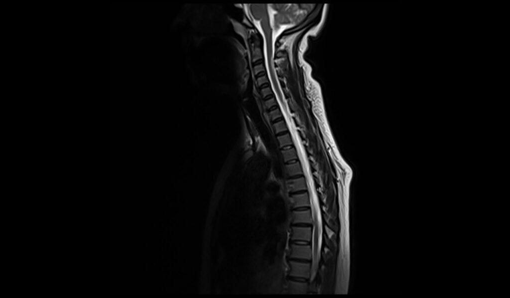
MRI T2 Appearance of Nitrous Oxide Toxicity in the Spinal Cord
T2-weighted MRI images are particularly sensitive to changes in water content within tissues and are thus useful in detecting edema and inflammation associated with nitrous oxide toxicity. In the spinal cord, T2-weighted images may show hyperintense (bright) signals in areas affected by nitrous oxide toxicity, indicating the presence of edema, demyelination, or gliosis. These hyperintense regions typically highlight the extent of the inflammatory response and the damage to the white matter tracts. T2 imaging is essential for identifying and evaluating the extent of the injury and the associated pathological changes in the spinal cord.
MRI STIR Appearance of Nitrous Oxide Toxicity in the Spinal Cord
Short Tau Inversion Recovery (STIR) sequences are highly sensitive to the presence of edema and can effectively suppress fat signals to provide clearer images of inflammatory processes. In the case of nitrous oxide toxicity, STIR images of the spinal cord may reveal pronounced hyperintensity in affected areas, highlighting the presence of significant edema and inflammation. These hyperintense signals are indicative of the acute phase of injury, where the inflammatory response is most active. STIR imaging is particularly useful for assessing the severity and distribution of the inflammatory changes in the spinal cord due to nitrous oxide toxicity.
T2 sagittal image of cervical spine shows Nitrous Oxide Toxicity in the Spinal Cord

T1 sagittal image of cervical spine shows Nitrous Oxide Toxicity in the Spinal Cord

STIR sagittal image of cervical spine shows Nitrous Oxide Toxicity in the Spinal Cord

T2 TSE axial image of image of cervical spine shows Nitrous Oxide Toxicity in the Spinal Cord


T1 TSE axial image of cervical spine shows Nitrous Oxide Toxicity in the Spinal Cord


References
- Safari, A., Emadi, F., Jamali, E., & Borhani-Haghighi, A. (2013). Clinical and MRI manifestations of nitrous oxide induced vitamin B12 deficiency: A case report. Iranian Journal of Neurology, 12(3), 111–113. https://www.ncbi.nlm.nih.gov/pmc/articles/PMC3829298/
- Vael, L., Van Walleghem, P., & Özsarlak, Ö. (2021). MRI of nitrous oxide-related subacute cervical myelopathy. Journal of the Belgian Society of Radiology, 105(1), 22. https://doi.org/10.5334/jbsr.2347
- Yuan, J. L., Wang, S. K., Jiang, T., & Hu, W. L. (2017). Nitrous oxide induced subacute combined degeneration with longitudinally extensive myelopathy with inverted V-sign on spinal MRI: a case report and literature review. BMC Neurology, 17, Article 222. https://doi.org/10.1186/s12883-017-0990-7