MRI herpes simplex encephalitis
Herpes simplex encephalitis (HSE) is a serious and potentially life-threatening condition characterized by inflammation of the brain (encephalitis) caused by the herpes simplex virus (HSV). This condition is relatively rare but can be severe, primarily affecting the temporal lobes of the brain.
Causes
Herpes simplex encephalitis is primarily caused by two strains of the herpes simplex virus:
- HSV-1: This is the most common cause of herpes simplex encephalitis and is typically associated with cold sores around the mouth. However, when it infects the brain, it can lead to encephalitis.
- HSV-2: While commonly associated with genital herpes, it can also cause encephalitis, although it’s less common for it to affect the brain compared to HSV-1.
The virus usually reaches the brain by traveling along nerve fibers, such as the olfactory nerves, or by spreading from an infection in another part of the body. In many cases, HSE arises when the virus reactivates from a latent state in a person who has previously been infected.
Symptoms
The symptoms of herpes simplex encephalitis can develop quickly, and early treatment is critical for a better outcome. Symptoms can vary but typically include:
- Fever: A sudden fever that is not associated with a common illness.
- Headache: Severe headache that can be persistent.
- Neurological issues: This can include confusion, seizures, personality changes, or hallucinations.
- Sensitivity to light: Known as photophobia.
- Stiff neck and back: Symptoms similar to those seen in meningitis.
- Altered consciousness: Ranging from mild disorientation to profound coma.
Diagnosis
Early and accurate diagnosis of herpes simplex encephalitis is critical for effective treatment. Diagnostic methods include:
- Lumbar puncture (spinal tap): This procedure involves collecting cerebrospinal fluid (CSF) to detect signs of inflammation and the presence of HSV DNA through polymerase chain reaction (PCR) testing, which is highly sensitive and specific for HSV.
- MRI (Magnetic Resonance Imaging): MRI scans can show abnormalities in the brain, particularly in the temporal lobes, which are commonly affected in HSE.
- EEG (Electroencephalogram): This test measures electrical activity in the brain and can show specific patterns that suggest encephalitis.
- Blood tests: These can help rule out other causes of the symptoms and assess the overall health and immune status of the patient.
Treatment
Treatment for herpes simplex encephalitis must be initiated quickly to reduce the risk of long-term neurological damage or death. Treatment typically includes:
- Antiviral Medications: Intravenous acyclovir is the standard treatment for herpes simplex encephalitis. This drug helps to reduce the activity of the virus but needs to be administered as early as possible.
- Steroids: Sometimes used to reduce brain swelling.
- Anticonvulsants: For patients experiencing seizures.
MRI appearance of herpes simplex encephalitis
T1-Weighted Imaging
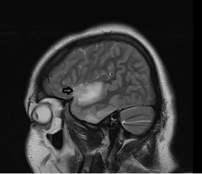
- In the early stages of HSE, there may be slight hypointensity or normal signal in affected areas. Later stages might show more pronounced hypointensity due to tissue destruction and necrosis.
T2-Weighted and FLAIR Imaging
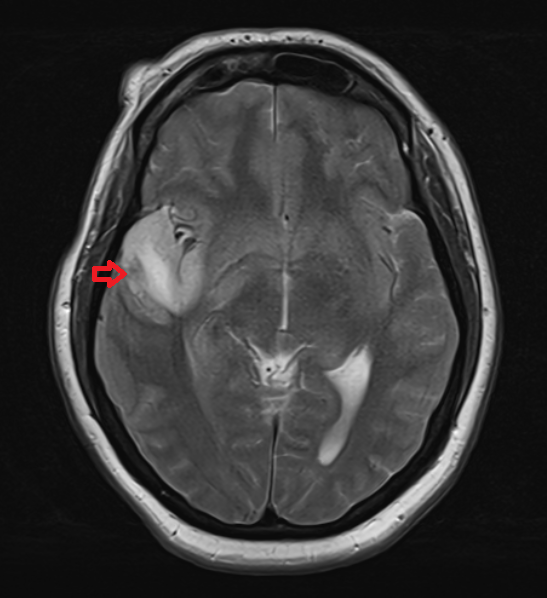
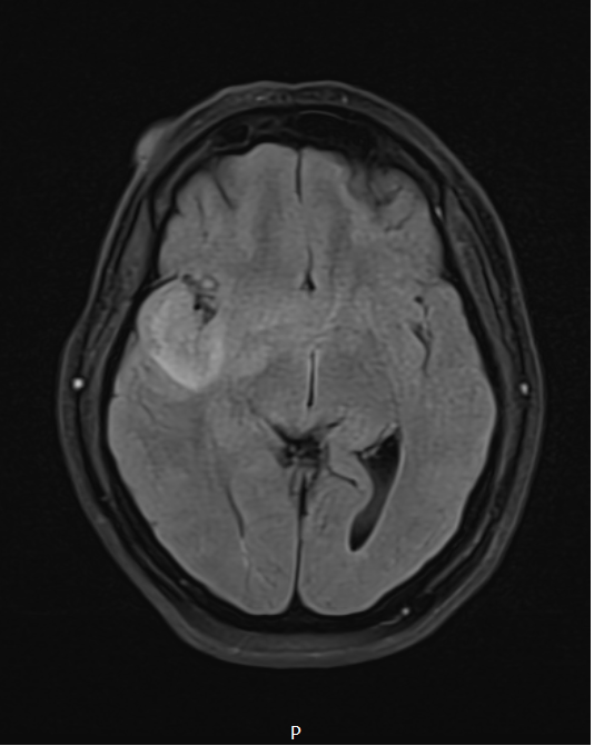
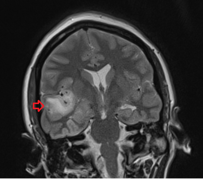
- These sequences show hyperintense signals in the areas of inflammation and edema. FLAIR imaging is particularly sensitive to detecting changes in the white matter and cortical regions.
Diffusion-Weighted Imaging (DWI) and ADC
- DWI (b0, b1000): Shows hyperintense signals in affected areas, indicating restricted diffusion due to cytotoxic edema.
- ADC: Areas that appear hyperintense on DWI typically exhibit hypointensity on ADC maps, confirming restricted diffusion.
Post-Contrast T1 Imaging
May show meningeal enhancement after contrast administration due to inflammation, although significant parenchymal enhancement is uncommon. When present, it might indicate more severe inflammation and breakdown of the blood-brain barrier.
T2 axial image shows herpes simplex encephalitis
FLAIR axial image shows herpes simplex encephalitis
T1 coronal image shows herpes simplex encephalitis

T2 sagittal image shows herpes simplex encephalitis
T2 coronal image shows herpes simplex encephalitis
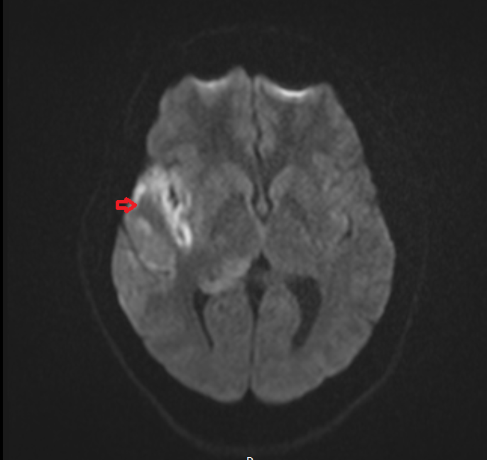
DWI b0 image shows herpes simplex encephalitis
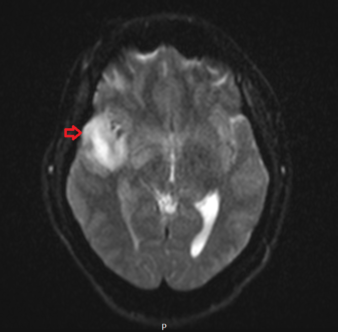
DWI b1000 image shows herpes simplex encephalitis
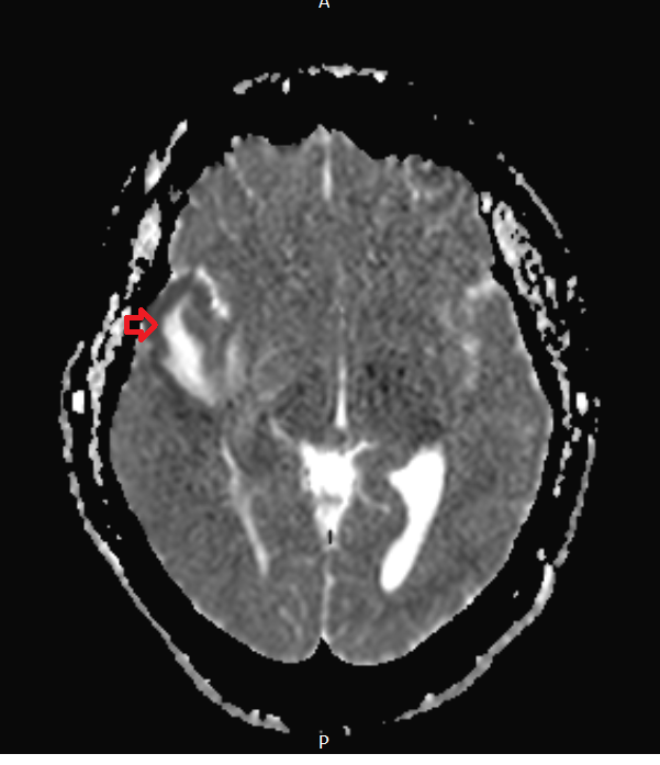
DWI ADC map image shows herpes simplex encephalitis
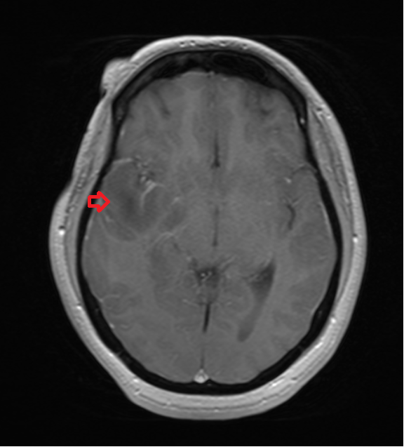
T1 post contrast axial image herpes simplex encephalitis
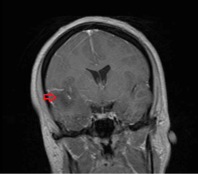
T1 post contrast coronal image herpes simplex encephalitis
Case study 2
T2 axial image shows herpes simplex encephalitis

T2 sagittal image shows herpes simplex encephalitis

FLAIR image shows herpes simplex encephalitis

T1 post contrast coronal image shows herpes simplex encephalitis

T1 post contrast axial image shows herpes simplex encephalitis

References
- Jayaraman, K., Rangasami, R. & Chandrasekharan, A., 2018. Magnetic Resonance Imaging Findings in Viral Encephalitis: A Pictorial Essay. Journal of Neuroscience in Rural Practice, 9(4), pp.556-560. Available at: https://www.ncbi.nlm.nih.gov/pmc/articles/PMC6126294/
- Sarton, B., Jaquet, P., Belkacemi, D., de Montmollin, E., Bonneville, F., Sazio, C., Frérou, A., Conrad, M., Daubin, D., Chabanne, R., Argaud, L., Dailler, F., Brulé, N., Lerolle, N., Maestraggi, Q., Marechal, J., Bailly, P., Razazi, K., Mateos, F., Guidet, B., Levrat, A., Susset, V., Lautrette, A., Mira, J.P., El Kalioubie, A., Robert, A., Massri, A., Albucher, J.F., Olivot, J.M., Conil, J.M., Boudma, L., Timsit, J.F., Sonneville, R., Silva, S., for the ENCEPHALITICA Consortium, 2021. Assessment of Magnetic Resonance Imaging Changes and Functional Outcomes Among Adults With Severe Herpes Simplex EncephalitisJAMA Network Open, 4(7), e2114328. Available at: https://doi.org/10.1001/jamanetworkopen.2021.14328
- Leonard, J.R., Moran, C.J., Cross, D.T., III, Wippold, F.J., II, Schlesinger, Y. and Storch, G.A., 2000. MR imaging of herpes simplex type I encephalitis in infants and young children: a separate pattern of findings. American Journal of Roentgenology, [online] 174(6). Available at: https://doi.org/10.2214/ajr.174.6.1741651
- McGrath, N., Anderson, N.E., Croxson, M.C., & Powell, K.F. (year). Herpes simplex encephalitis treated with acyclovir: diagnosis and long term outcome. Journal of Neurology, Neurosurgery & Psychiatry, 63(3). Available at: https://jnnp.bmj.com/content/63/3/321