MRI Dysplastic cerebellar gangliocytoma (Lhermitte-Duclos disease)
MRI Dysplastic cerebellar gangliocytoma, also known as Lhermitte-Duclos disease (LDD), is a rare, slow-growing, benign brain tumor that predominantly affects the cerebellum. It is characterized by an abnormal growth of large, dysplastic ganglion cells, replacing the normal granular layer of the cerebellum. This condition was first described by Lhermitte and Duclos in 1920 and is often associated with Cowden syndrome, a genetic disorder that increases the risk of developing multiple tumor types.
Cause
The exact cause of Lhermitte-Duclos Disease is not fully understood. However, it is often associated with Cowden syndrome, a genetic disorder characterized by multiple noncancerous, tumor-like growths and an increased risk of certain cancers.
Symptoms
Symptoms of Lhermitte-Duclos disease typically arise from increased intracranial pressure or mass effect due to the tumor’s growth, which can compress adjacent structures in the brain. Common symptoms include:
- Headaches
- Nausea and vomiting
- Visual disturbances
- Difficulty with coordination and balance (ataxia)
- Dizziness
- Tinnitus or hearing loss
- Cognitive impairments
Diagnosis
Diagnosis of Lhermitte-Duclos Disease involves a combination of clinical evaluation and imaging studies. Magnetic Resonance Imaging (MRI) is crucial for diagnosis, as it provides detailed images of the brain’s structures, helping to identify the characteristic appearance of the tumor in the cerebellum. The tumor typically appears as a non-enhancing mass with a striated pattern, often referred to as a “tiger-striped” appearance on MRI scans.
Treatment
The treatment of Lhermitte-Duclos disease depends on the size of the tumor and its location.
- Surgical Removal: The primary treatment method, aiming to reduce intracranial pressure and alleviate symptoms. Complete removal can be challenging due to the tumor’s size and location.
- Monitoring: In cases where the tumor is not causing significant symptoms or its growth is very slow, careful monitoring with regular MRI scans may be recommended.
MRI appearance of Lhermitte-Duclos disease
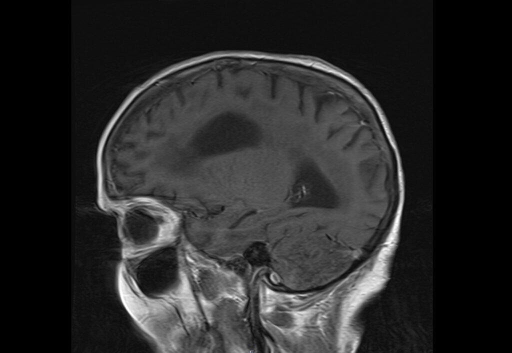
T1-Weighted Images
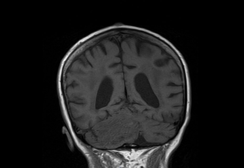
- On T1-weighted MRI, Lhermitte-Duclos disease typically appears as hypointense (darker than surrounding brain tissue) or isointense relative to normal cerebellar white matter. This reflects the dense fibrous tissue and abnormal large ganglion cells replacing the normal cerebellar architecture.
T2-Weighted Images
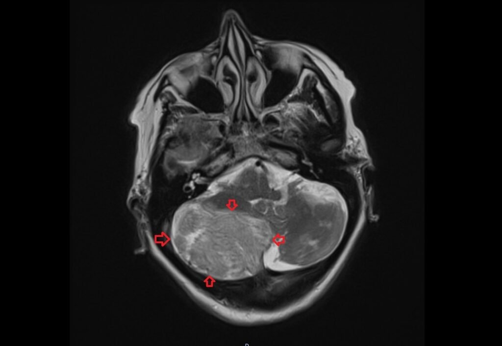
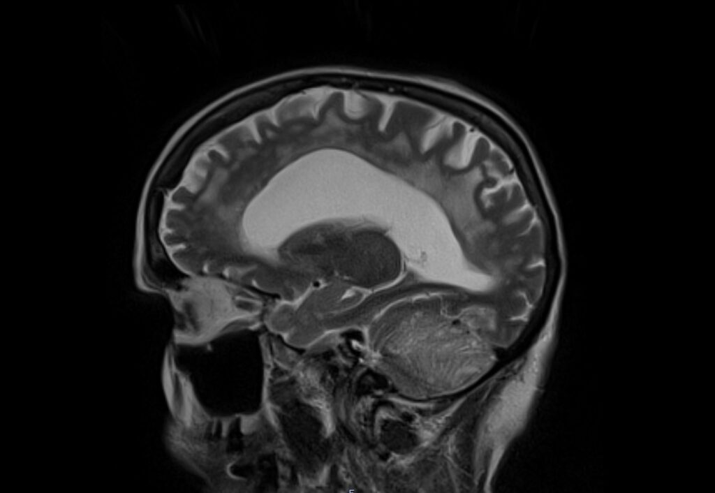
- T2-weighted imaging is particularly useful for visualizing Lhermitte-Duclos disease. The tumor typically displays a striated or “tiger-striped” appearance, which is very characteristic. This pattern results from alternating bands of high and low signal intensity. The high signal intensity areas correspond to myxoid changes and abnormal ganglion cells, whereas the low signal areas reflect fibrous bands.
3. FLAIR (Fluid Attenuated Inversion Recovery)
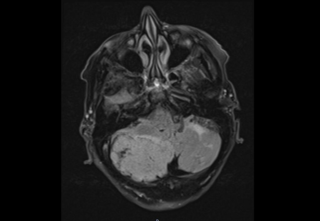
- On FLAIR images, Lhermitte-Duclos disease also appears as hyperintense with the same striped pattern noted on T2-weighted images. FLAIR is particularly helpful in suppressing the cerebrospinal fluid signal, enhancing the contrast between the lesion and the cerebellar fluid, which aids in delineating the extent of the lesion.
4. DWI (Diffusion Weighted Imaging)
- Appearance: Typically, Lhermitte-Duclos disease shows no restricted diffusion on DWI, which suggests a lack of acute cell death or high cellularity that is often seen in more aggressive tumors.
- Details: The apparent diffusion coefficient (ADC) values are usually normal or elevated, indicating facilitated diffusion which is consistent with a benign and less dense cellular structure.
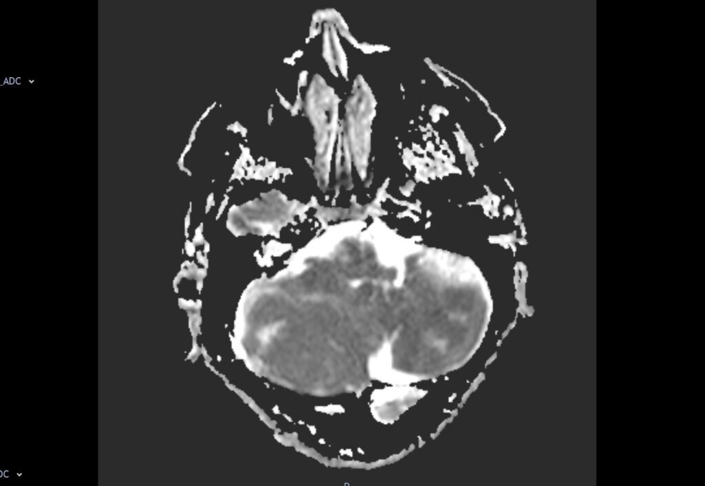
DWI and ADC
- DWI (Diffusion Weighted Imaging): Dysplastic cerebellar gangliocytoma typically shows no restricted diffusion on DWI, appearing isointense or slightly hyperintense compared to surrounding brain tissue.
- ADC (Apparent Diffusion Coefficient): On ADC maps, the lesion usually exhibits increased diffusion, reflecting the non-restrictive, expanded extracellular space characteristic of the tumor.
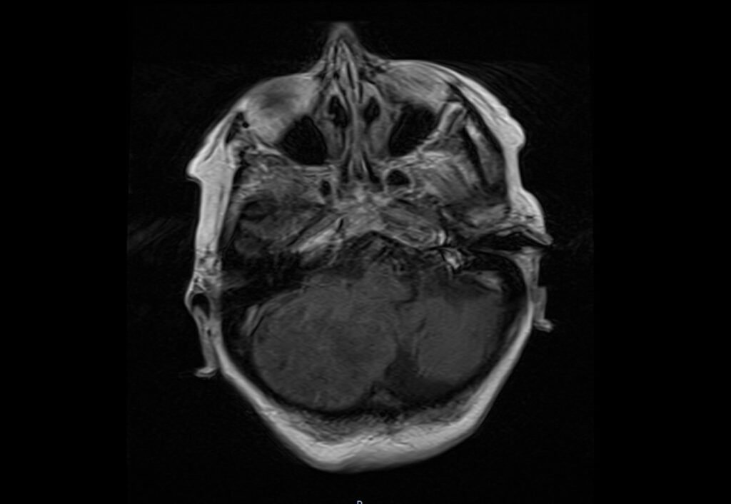
Post-Contrast T1
- After the administration of gadolinium-based contrast agents, there is generally no significant enhancement in Lhermitte-Duclos disease, which helps to differentiate it from other neoplastic lesions that might enhance.
T2 axial image shows Dysplastic cerebellar gangliocytoma (Lhermitte-Duclos disease)
FLAIR axial image shows Dysplastic cerebellar gangliocytoma (Lhermitte-Duclos disease)
T1 coronal image shows Dysplastic cerebellar gangliocytoma (Lhermitte-Duclos disease)
T2 sagittal image shows Dysplastic cerebellar gangliocytoma (Lhermitte-Duclos disease)
DWI b0 image shows Dysplastic cerebellar gangliocytoma (Lhermitte-Duclos disease)
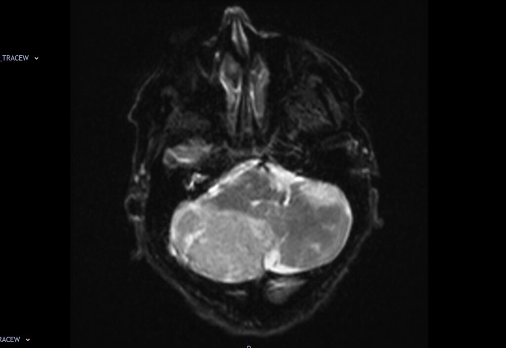
DWI b1000 image shows Dysplastic cerebellar gangliocytoma (Lhermitte-Duclos disease)

DWI ADC map image shows Dysplastic cerebellar gangliocytoma (Lhermitte-Duclos disease)
T1 post contrast axial image shows Dysplastic cerebellar gangliocytoma (Lhermitte-Duclos disease)
T1 post contrast sagittal image shows Dysplastic cerebellar gangliocytoma (Lhermitte-Duclos disease)
References
- Wan, X., Chu, H., Chen, W., & Wang, F. (2021). Dysplastic cerebellar gangliocytoma: a description of two cases. Quantitative Imaging in Medicine and Surgery, 11(11), 4695–4699. https://doi.org/10.21037/qims-20-627
- Puiseux, C., Bretonnier, M., Proisy, M., Chappé, C., Denizeau, P., & Riffaud, L. (2021). Dysplastic gangliocytoma of the cerebellum (Lhermitte-Duclos disease) presenting as a prenatally heterotopic hamartoma. Child’s Nervous System, 37(2021), 1017–1020. doi:10.1007/s00381-020-04826-2
- Zhang, H.-w., Zhang, Y.-q., Liu, X.-l., Mo, Y.-q., Lei, Y., Lin, F., & Feng, Y.-n. (2022). MR imaging features of Lhermitte–Duclos disease: Case reports and literature review. Medicine (Baltimore), 101(4), e28667. https://doi.org/10.1097/MD.0000000000028667
- Joo, G., & Doumanian, J. (2020). Radiographic Findings of Dysplastic Cerebellar Gangliocytoma (Lhermitte-Duclos Disease) in a Woman with Cowden Syndrome: A Case Study and Literature Review. Journal of Radiology Case Reports, 14(3), 1–6. https://doi.org/10.3941/jrcr.v14i3.3814
- Lakhani, D. A., & Boo, S. (2022, December 13). Images in Radiology: Lhermitte-Duclos Disease. Radiology, 307(1). Retrieved from https://doi.org/10.1148/radiol.221979