Cervical cancer MRI
Cervical cancer is a type of cancer that occurs in the cells of the cervix, which is the lower part of the uterus that connects to the vagina. It’s one of the significant health issues facing women globally but can often be successfully treated, especially when detected early.
Causes
The primary cause of cervical cancer is a persistent infection of certain types of human papillomavirus (HPV). HPV is a common virus that is transmitted through sexual contact. There are many types of HPV, but only some of them cause cervical cancer. Other factors can increase the risk of developing cervical cancer, including:
- Smoking
- Having a weakened immune system
- Long-term use of oral contraceptives
- Having many sexual partners or becoming sexually active early
- Other sexually transmitted infections (STIs)
Symptoms
In its early stages, cervical cancer typically does not produce any symptoms. As the cancer progresses, symptoms may include:
- Vaginal bleeding after intercourse, between periods, or after menopause
- Watery, bloody vaginal discharge that may be heavy and have a foul odor
- Pelvic pain or pain during intercourse
Diagnosis
Cervical cancer is typically diagnosed through a series of steps:
- Pap smear test: This test involves collecting cells from the cervix, which are then examined under a microscope to find precancerous or cancerous cells.
- HPV DNA test: This test checks for the presence of the virus that can lead to cervical changes.
- Biopsy: A small sample of tissue is taken from the cervix and examined under a microscope to check for cancer cells.
Treatment
Treatment for cervical cancer depends on several factors, including the stage of the cancer, overall health, and personal preferences. Common treatment options include:
- Surgery: This can range from a simple procedure that removes precancerous cells to more extensive operations that may involve removing the cervix, part of the vagina, and possibly the uterus (hysterectomy).
- Radiation therapy: This uses high-energy rays to kill cancer cells. It can be given externally or internally (brachytherapy).
- Chemotherapy: This involves using drugs to kill cancer cells, usually given intravenously. It’s often used alongside radiation therapy.
- Targeted therapy: Drugs or other substances that target specific cancer cells without affecting normal cells.
- Immunotherapy: Uses the body’s immune system to fight cancer. It may be an option if cervical cancer has advanced or returned after treatment.
Prevention
Preventive measures include:
- HPV vaccination: Available for both boys and girls, this vaccine protects against the types of HPV most likely to cause cervical cancer.
- Regular screening tests: Pap tests and HPV tests help detect cervical precancers and cancers early.
MRI Appearance of Cervical cancer
T2-weighted: T2-weighted images provide excellent contrast between the high-signal intensity of the tumor and the low-signal intensity of the surrounding fibrous stroma. Cervical cancers are usually seen as higher signal intensity relative to the low signal cervical stroma, making this sequence particularly useful for assessing tumor size and local spread.
DWI (Diffusion Weighted Imaging) and ADC (Apparent Diffusion Coefficient):
- B0: This initial sequence in DWI might not specifically highlight the tumor but sets the baseline for diffusion measurement.
- B1000: At a high b-value like B1000, areas of restricted diffusion (common in malignant tissues like cervical cancer) will appear very bright. This is because cancer cells typically are densely packed, restricting the diffusion of water molecules.
- ADC map: This map calculates the diffusion rate of water molecules within tissue. Cervical cancers typically show low ADC values due to high cellularity which restricts water movement. Areas of the tumor on the ADC map will appear darker compared to the surrounding tissue.
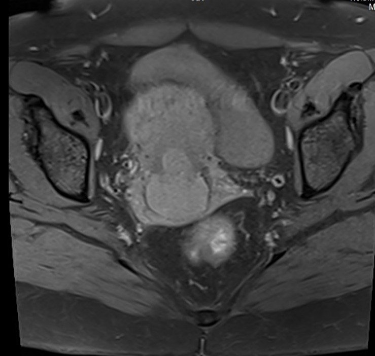
T1-weighted Fat-Saturated: This sequence helps in better visualization of the tumor against the suppressed fat signal. Cervical cancer typically appears as a mass that is iso-intense to the surrounding muscle or slightly hypo-intense.
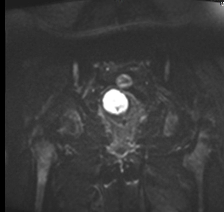
STIR (Short TI Inversion Recovery): STIR is useful for suppressing fat signals and highlighting edema and inflammation, which is common in tumor-involved areas. In the context of cervical cancer, STIR images are particularly valuable for assessing the spread of the disease to lymph nodes and other soft tissues beyond the cervix.
T2 TSE sagittal image shows Cervical cancer

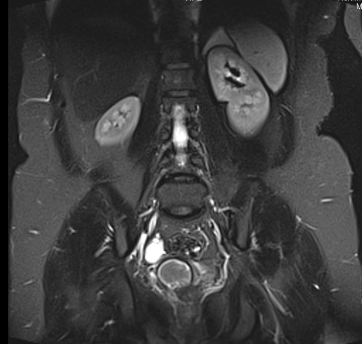
T2 coronal oblique image shows Cervical cancer
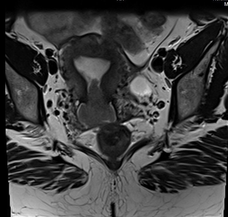
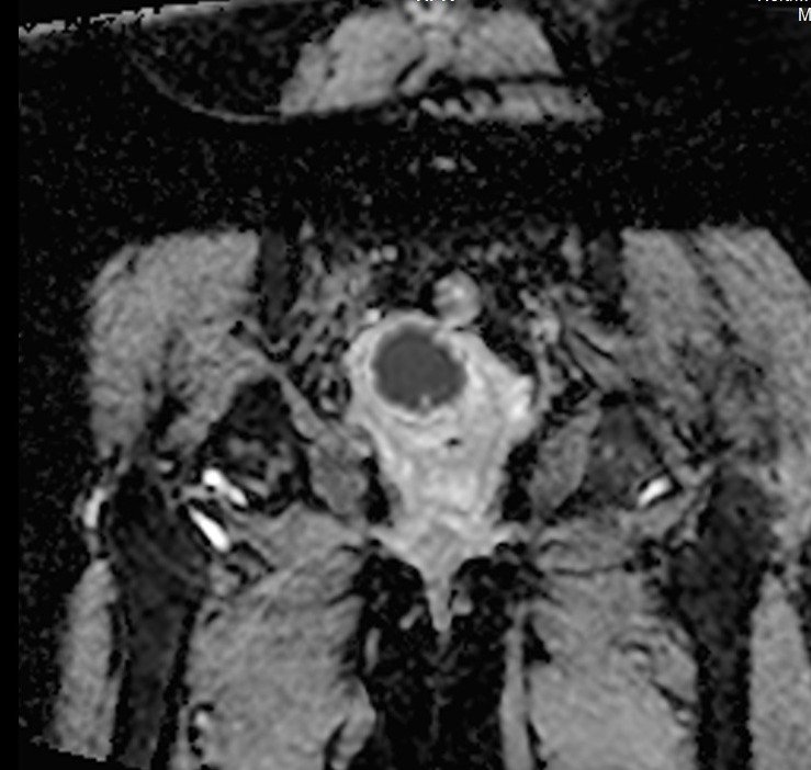
T2 axial oblique image shows Cervical cancer

T1 fat saturated coronal oblique image shows Cervical cancer
DWI epi b0 axial oblique image shows Cervical cancer
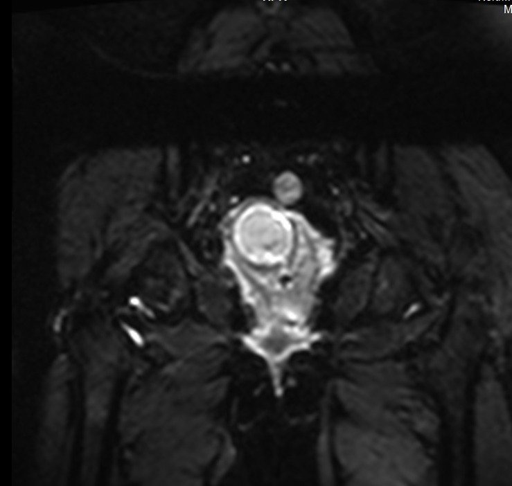
DWI epi b1000 axial oblique image shows Cervical cancer
DWI ADC axial oblique image shows Cervical cancer
STIR coronal image shows Cervical cancer
References
- Mansoori, B., Khatri, G., Rivera-Colón, G., Albuquerque, K., Lea, J., & Pinho, D. F. (2020, June 17). Multimodality Imaging of Uterine Cervical Malignancies. American Journal of Roentgenology, 215(2). https://doi.org/10.2214/AJR.19.21941
- Avesani, G., Perazzolo, A., Amerighi, A., Celli, V., Panico, C., Sala, E., & Gui, B. (2023). The Utility of Contrast-Enhanced Magnetic Resonance Imaging in Uterine Cervical Cancer: A Systematic Review. Life (Basel), 13(6), 1368. https://doi.org/10.3390/life13061368
- Otero-García, M. M., Mesa-Álvarez, A., Nikolic, O., Blanco-Lobato, P., Basta-Nikolic, M., Menéndez de Llano-Ortega, R., Paredes-Velázquez, L., Nikolic, N., & Szewczyk-Bieda, M. (2019). Role of MRI in staging and follow-up of endometrial and cervical cancer: pitfalls and mimickers. Insights into Imaging, 10, 19.
- Balleyguier, C., Sala, E., Da Cunha, T., Bergman, A., Brkljacic, B., Danza, F., Forstner, R., Hamm, B., Kubik-Huch, R., Lopez, C., Manfredi, R., McHugo, J., Oleaga, L., Togashi, K., & Kinkel, K. (2011). Staging of uterine cervical cancer with MRI: guidelines of the European Society of Urogenital Radiology. European Radiology, 21, 1102–1110.