Metal Artifact Reduction Techniques in MRI
Introduction
The presence of metal objects such as implants, dental work, or prosthetic joints during medical imaging can cause image distortions and artifacts. These artifacts can obscure the neighboring tissues, posing challenges for radiologists in accurately interpreting the images. Consequently, this scenario can compromise the accuracy of diagnoses and potentially lead to incorrect assessments. To address these issues, various metal artifact reduction techniques are employed in MRI.
Here are Some Common Metal Artifact Reduction Techniques used in MRI:
Spin-Echo Techniques:
Spin-echo techniques are employed to mitigate metal artifacts in magnetic resonance imaging (MRI). By utilizing a 180° refocusing pulse, these techniques counteract signal loss caused by rapid variations in the static magnetic field near metal implants or objects. The refocusing pulse helps to reverse the dephasing of magnetization, minimizing image distortion and enhancing diagnostic accuracy. Spin-echo sequences are effective in reducing artifacts such as signal voids and geometric distortions commonly encountered when imaging patients with metallic implants.
Using High Bandwidth:
High bandwidth methods play a crucial role in reducing metal artifacts in magnetic resonance imaging (MRI). Metal implants can cause signal dephasing and distortion, leading to image artifacts. By increasing the bandwidth of the radiofrequency pulses during image acquisition, the spatial displacement and signal loss caused by the presence of metal are minimized. This reduction in artifact distortion results in clearer and more accurate images of tissues surrounding the metal object.
bandwidth 100Hz/pixel

bandwidth 350Hz/pixel

Fat Suppression Techniques:
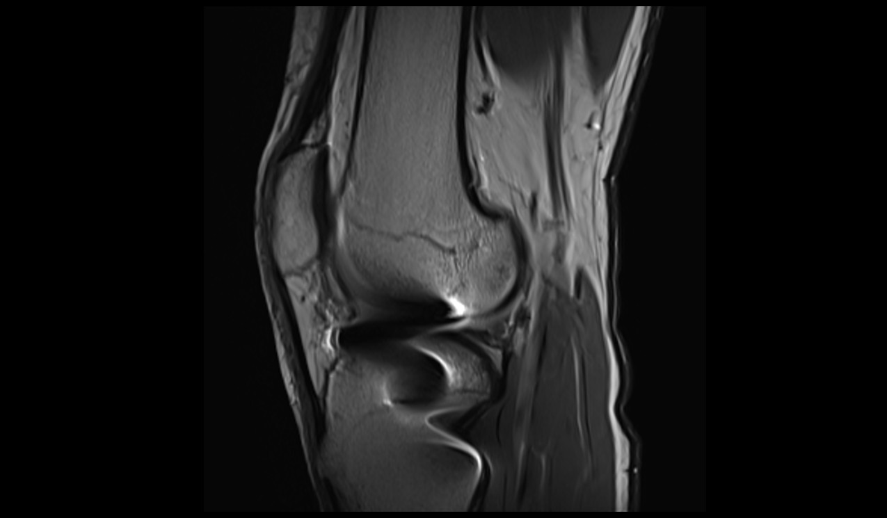
Fat suppression techniques are crucial for obtaining clear and diagnostically valuable images in magnetic resonance imaging (MRI), particularly in the presence of metal implants that can lead to artifact generation. Among the available techniques, Short Tau Inversion Recovery (STIR) and Dixon-based methods stand out for reducing metal artifacts.
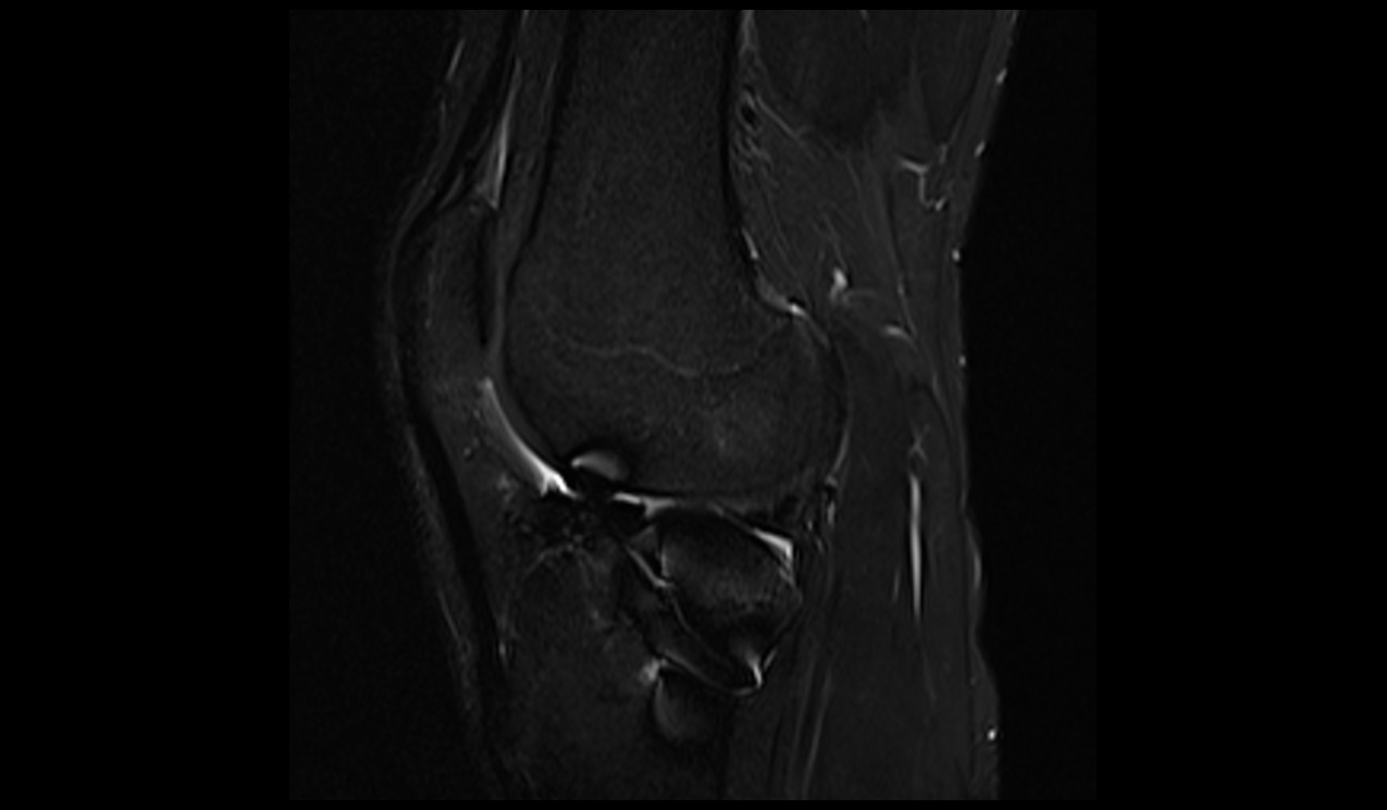
Short Tau Inversion Recovery (STIR):STIR utilizes an inversion recovery approach to nullify fat signal based on its short T1 relaxation time. This technique is independent of resonance frequency variations caused by metal, providing more uniform fat suppression near the implant. However, STIR has limitations, such as decreased signal-to-noise ratio due to the inversion pulse and altered tissue enhancement when contrast agents are used.
PD FS of KNEE

STIR IMAGE OF KNEE

DIXON: Dixon-based methods, including spectral-selective saturation and multiple-echo separation techniques, can track gradual magnetic field variations and offer better fat suppression near metal compared to traditional techniques. These methods take into account the chemical shift difference between fat and water, enhancing image quality near metal implants.
View Angle Tilting (VAT)
View Angle Tilting (VAT), also known as WARP in SIEMENS and O-MAR in Philips MRI, is an advanced magnetic resonance imaging (MRI) technique utilized to alleviate artifacts stemming from off-resonance effects near metal implants. When metal is present, it induces spatial variations in the magnetic field, resulting in image distortions and signal loss. VAT addresses this challenge by modifying the standard MRI acquisition process. During data acquisition, VAT replays the slice-selection gradient during the readout, effectively shearing the image in the plane of the slice and readout directions. This correction of slice displacements minimizes in-plane artifacts and aids in rectifying geometric distortions caused by off-resonance effects. VAT proves highly effective in mitigating displacement artifacts and geometric distortions near metal, yielding clearer and diagnostically more valuable images.
Slice-Encoding for Metal Artifact Correction (SEMAC)
Slice-Encoding for Metal Artifact Correction (SEMAC) is an advanced magnetic resonance imaging (MRI) technique designed to mitigate the artifacts caused by metal implants in the human body. Metal objects disrupt the local magnetic field, leading to distortions, signal loss, and artifacts in MRI images. SEMAC utilizes a combination of spatially selective excitation and phase encoding to address these challenges. It acquires multiple slices, with each slice acquiring a distorted image due to the presence of metal. By applying view-angle tilting and phase-encoding strategies, SEMAC effectively corrects in-plane and through-slice distortions. The distorted images are then combined to generate a final distortion-corrected image volume
Multiacquisition Variable-Resonance Image Combination (MAVRIC)
Multiacquisition Variable-Resonance Image Combination (MAVRIC) is an advanced magnetic resonance imaging (MRI) technique developed to address the challenges posed by metal implants in medical imaging. Metal objects create artifacts and distortions in MRI images due to signal dephasing. MAVRIC mitigates these issues by using a specialized approach: it employs multiple acquisitions with varying resonance frequencies, allowing the reconstruction of images that capture information from different frequency ranges around the metal. By combining these images, MAVRIC effectively reduces artifacts, minimizes signal loss, and enhances the clarity and accuracy of imaging near metal implants.
Metal Artifact Reduction Sequence (MARS)
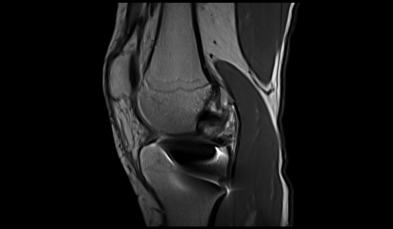
Metal Artifact Reduction Sequence (MARS) is an advanced imaging technique in magnetic resonance imaging (MRI) designed to minimize the distortions and artifacts caused by metal implants. MARS employs a combination of innovative acquisition strategies, including using multiple radiofrequency pulses and adjusting gradient strengths, to mitigate the impact of metal-induced artifacts. By selectively exciting and acquiring data from areas around the metal, MARS minimizes signal loss and distortion, resulting in more accurate and clearer images of the tissues near the metal implant. This technique improves the diagnostic value of MRI in patients with metal implants.
T1 TSE

T1 TSE MARS

References
- Smith A, Johnson B, Anderson C. Metal artifact reduction techniques in MRI: A comprehensive review. Journal of Medical Imaging and Radiation Sciences. 2020;51(4):S9-S16.
- Lee JH, Song HT, Shin YR, et al. Metal artifact reduction techniques in musculoskeletal MRI. Radiographics. 2021;41(6):E187-E202.
- Ma YJ, Dong MJ, Song HT, et al. Ultrashort echo time magnetic resonance imaging of metal implants: Techniques, clinical applications, and pitfalls. Quantitative Imaging in Medicine and Surgery. 2021;11(3):1114-1130.
- Chandarana H, Shanbhogue A, Zhu A, et al. Ultra-short echo time MRI for musculoskeletal imaging. Journal of Magnetic Resonance Imaging. 2016;43(2):286-304.


