MRI Lipoma of the Conus
Lipoma of the conus is a rare condition referring to a benign tumor composed of adipose (fat) tissue located in the conus medullaris, the lower end of the spinal cord. This condition falls under the category of spinal lipomas, which are one of the most common forms of spinal cord tumors in children, but they can also be found in adults.
Symptoms
The symptoms of lipoma of the conus can vary greatly depending on the size and location of the tumor. Common symptoms include:
- Back Pain: This is a frequent symptom, particularly if the lipoma is large or is causing nerve compression.
- Neurological Symptoms: These can include numbness, weakness, or tingling in the lower limbs, reflecting the involvement of the spinal cord and nerves.
- Bladder and Bowel Dysfunction: In some cases, the lipoma can interfere with the nerves that control bladder and bowel functions, leading to incontinence or difficulty in urination and defecation.
Treatment
Treatment of a lipoma of the conus medullaris depends on several factors, including the size of the lipoma, the severity of symptoms, and the patient’s overall health. Options include:
Observation: In cases where the lipoma is small and not causing significant symptoms, regular monitoring may be recommended.
Surgery: This is the primary treatment for symptomatic lipomas. The goal is to remove as much of the lipoma as safely possible to relieve pressure on the spinal cord and nerves. Complete removal can be challenging and risky, given the involvement of vital neural structures.
Symptom Management: This includes pain management, physical therapy, and, in some cases, managing bowel or bladder dysfunction.
MRI Appearance of conus medullaris lipoma
T1-weighted images:
- Lipomas typically appear hyperintense (bright) on T1-weighted images because of the high fat content. The contrast between the lipoma and the surrounding structures is quite pronounced.
T2-weighted images:
- On T2-weighted images, the lipoma may also appear hyperintense, but the contrast with the surrounding cerebrospinal fluid (which is also bright on T2) might be less apparent.
T1-weighted images with fat suppression (T1 Fat Sat):
- This sequence is crucial for confirming the fatty nature of the lesion. The lipoma, which appears bright on regular T1 images, will lose signal and become dark on fat-suppressed images, distinguishing it from other types of lesions.
Short Tau Inversion Recovery (STIR):
- In this sequence, fat signal is suppressed, making the lipoma appear dark. This is similar to fat-suppressed T1, but STIR is more sensitive to water content and can highlight edema or inflammation in adjacent tissues.
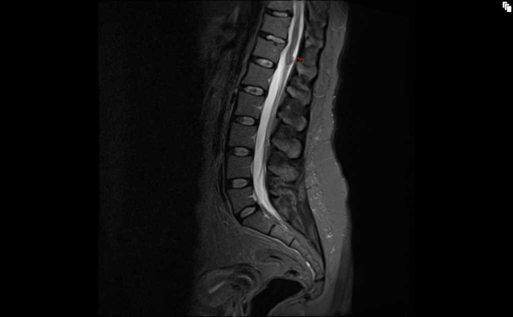
T2 sagittal image shows conus medullaris lipoma

T1 TSE sagittal image shows conus medullaris lipoma

STIR sagittal image shows conus medullaris lipoma
T2 TSE axial image shows conus medullaris lipoma
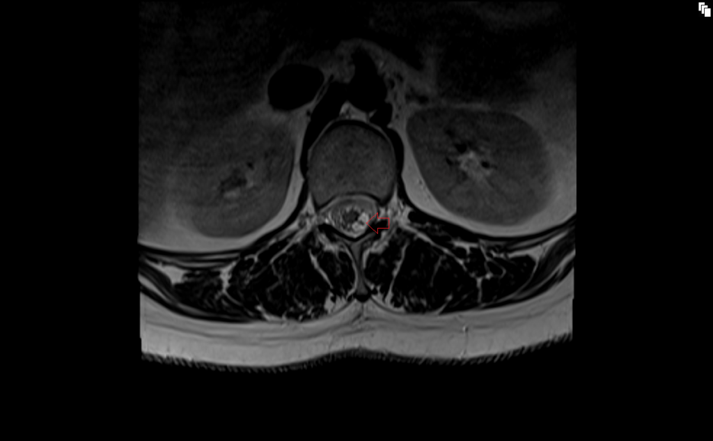
T1 TSE axial image shows conus medullaris lipoma

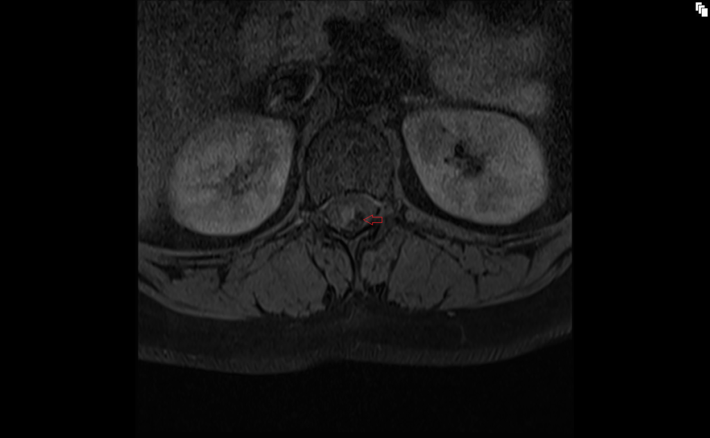
T1 TSE fat sat axial image shows conus medullaris lipoma
References
- Gupta, A., Bansal, K., Kalidindi, K. K. V., Bhargava, A., & Verma, A. (2021). Intradural Conus Medullaris Lipoma With Neurological Deficit: A Rare Occurrence. Cureus, 13(3), e14053. doi: 10.7759/cureus.14053.
- Coran, A., Ortolan, P., Attar, S., Alberioli, E., Perissinotto, E., Tosi, A. L., Montesco, M. C., Rossi, C. R., Tropea, S., Rastrelli, M., & Stramare, R. (2017). Magnetic Resonance Imaging Assessment of Lipomatous Soft-tissue Tumors. In Vivo, 31(3), 387–395. doi: 10.21873/invivo.11071
- Burt, A. M., & Huang, B. K. (2017). Imaging review of lipomatous musculoskeletal lesions. SICOT J, 3, 34. https://doi.org/10.1051/sicotj/2017015
- Patwardhan, V., Patanakar, T., Armao, D., & Mukherji, S. K. (June 2000). MR Imaging Findings of Intramedullary Lipomas. American Journal of Roentgenology, 174(6). https://doi.org/10.2214/ajr.174.6.1741792
- Komiyama, M., Hakuba, A., Inoue, Y., Yasui, T., Yagura, H., Baba, M., & Nishimura, S. (1987). Magnetic resonance imaging: Lumbosacral lipoma. Surgical Neurology, 28(4), 259-264.