Knee Synovitis MRI
Synovitis is the inflammation of the synovial membrane, which is a layer of connective tissue that lines the cavities of joints, tendon sheaths, and bursae (fluid-filled sacs). This membrane produces synovial fluid, which lubricates and nourishes the joints. Synovitis can occur in any joint but is most commonly found in the knees, wrists, elbows, and hips.
Causes
- Injury: Trauma to the joint can cause synovitis.
- Rheumatoid arthritis: An autoimmune condition where the body’s immune system attacks the synovium.
- Infection: Bacterial or viral infections can lead to joint inflammation.
- Gout: Accumulation of uric acid crystals in the joint.
- Osteoarthritis: Degenerative joint disease causing wear and tear of the cartilage.
Symptoms
- Pain: The affected joint is typically painful.
- Swelling: The joint may become swollen and appear larger than usual.
- Warmth: The joint may feel warm to the touch.
- Stiffness: Reduced range of motion and stiffness, especially after periods of inactivity.
- Redness: The skin over the joint may appear red.
- Fatigue: General tiredness and fatigue, especially in cases of rheumatoid arthritis.
Diagnosis
- Clinical Examination: A thorough physical examination to check for signs of inflammation.
- Medical History: Reviewing the patient’s medical history and symptoms.
- Imaging Tests:
- X-rays: To check for joint damage or deformities.
- Ultrasound: To detect inflammation and fluid in the joint.
- MRI: To get a detailed image of the joint structures.
- Laboratory Tests:
- Blood Tests: To check for markers of inflammation and autoimmune conditions (e.g., rheumatoid factor, anti-CCP antibodies, ESR, CRP).
- Joint Aspiration: Removing a sample of synovial fluid from the joint for analysis to check for infection, crystals, or inflammatory markers.
Treatment
Treatment for synovitis aims to reduce inflammation and pain, as well as address any underlying causes. Common treatments include:
- Cold and heat therapy: Applying ice packs to reduce swelling and heat to relieve stiffness.
- Medications: Nonsteroidal anti-inflammatory drugs (NSAIDs) to reduce pain and inflammation. In more severe cases, corticosteroids may be injected directly into the joint.
- Physical therapy: Exercises to improve range of motion and strengthen the muscles around the joint.
- Surgery: In severe cases, particularly where there is recurrent or chronic synovitis, surgical intervention might be necessary to remove inflamed tissue or correct joint abnormalities.
MRI Appearance of Knee Synovitis
MRI STIR Appearance of Knee Synovitis
- Short Tau Inversion Recovery (STIR) sequences in MRI are highly sensitive for detecting inflammation and fluid, making them ideal for identifying knee synovitis. In cases of synovitis, STIR images typically show hyperintense (bright) signals within the synovial membrane and joint cavity, indicating the presence of inflammation and increased fluid. This bright signal helps to distinguish areas of active inflammation from other tissues, providing a clear visualization of the extent and severity of synovitis. The STIR sequence is particularly useful in early detection and monitoring of inflammatory changes in the knee joint.
MRI T1 Appearance of Knee Synovitis
- T1-weighted MRI sequences provide excellent anatomical detail and are useful for assessing the structure of the knee joint. In the context of knee synovitis, T1 images may show a thickened synovial membrane with intermediate signal intensity compared to the surrounding tissues. While T1-weighted images are not as sensitive as STIR or T2 for detecting fluid and inflammation, they are valuable for identifying structural changes and chronic synovitis, which may appear as regions of synovial hypertrophy or fibrosis. These images help in assessing the overall joint anatomy and any associated degenerative changes.
MRI PD FS Appearance of Knee Synovitis
- Proton Density Fat-Suppressed (PD FS) sequences combine high anatomical detail with enhanced sensitivity to fluid, making them effective for evaluating knee synovitis. In cases of synovitis, PD FS images typically reveal hyperintense (bright) areas within the synovial membrane and joint space, similar to STIR sequences but with better contrast and resolution. The fat suppression technique enhances the visibility of fluid and inflammation by reducing the signal from fat, providing a clearer view of the inflamed synovial tissue. This sequence is particularly useful for detailed assessment of synovial inflammation and for planning targeted treatments.
MRI MEDIC Appearance of Knee Synovitis
- Multi-Echo Data Image Combination (MEDIC) sequences provide high-resolution images with excellent contrast, making them useful for detailed assessment of joint structures in knee synovitis. MEDIC sequences can reveal both the hyperintense signal of inflamed synovial tissue and the fine anatomical details of the joint. These sequences are particularly beneficial in evaluating complex cases of synovitis where detailed visualization of the synovial membrane and adjacent structures is required.
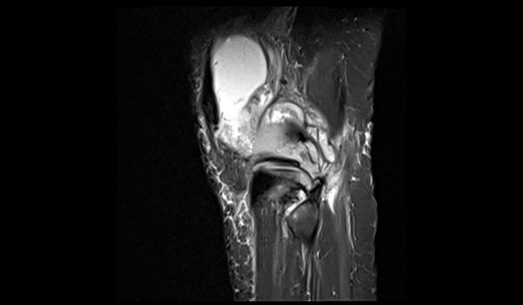
STIR sagittal image shows Knee Synovitis


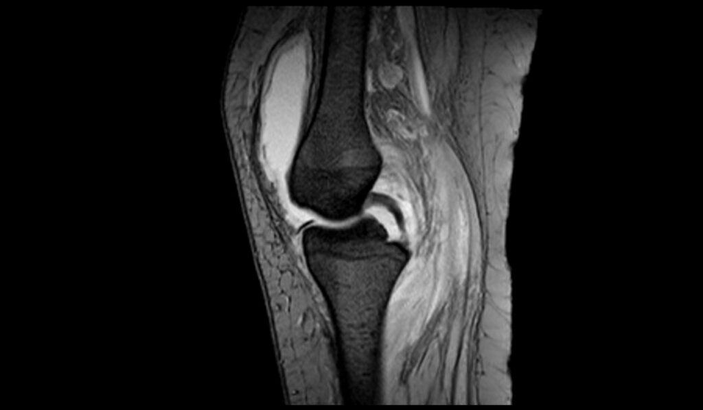
T1 sagittal image shows Baker's cyst rupture


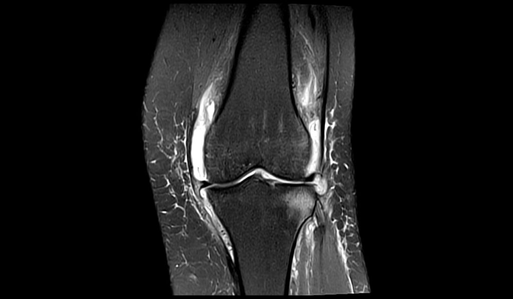
PD FS coronal image shows Baker's cyst rupture


PD FS axial image shows Baker's cyst rupture


MEDIC(T2*) sagittal image shows Baker's cyst rupture


References
- Burke, C. J., Alizai, H., Beltran, L., & Regatte, R. R. (2019). MRI of synovitis and joint fluid. Journal of Magnetic Resonance Imaging, 49(6), 1512–1527. https://doi.org/10.1002/jmri.26618
- Thoenen, J., MacKay, J. W., Sandford, H. J. C., Gold, G. E., & Kogan, F. (2021). Imaging of synovial inflammation in osteoarthritis, from the AJR special series on inflammation. American Journal of Roentgenology, 218(3).
- Huang, G.-S., Lee, C.-H., Chan, W. P., Chen, C.-Y., Yu, J. S., & Resnick, D. (2003). Localized nodular synovitis of the knee: MR imaging appearance and clinical correlates in 21 patients. American Journal of Roentgenology, 181(2).
- de Vries, B. A., Breda, S. J., Sveinsson, B., McWalter, E. J., Meuffels, D. E., Krestin, G. P., Hargreaves, B. A., Gold, G. E., & Oei, E. H. G. (2021). Detection of knee synovitis using non-contrast-enhanced qDESS compared with contrast-enhanced MRI. Arthritis Research & Therapy, 23, Article 55. https://doi.org/10.1186/s13075-021-02429-7