Knee Lymphoma MRI
Lymphoma is a type of cancer that originates in the lymphatic system, which is a crucial part of the body’s immune system. The lymphatic system includes the lymph nodes, spleen, thymus gland, and bone marrow. There are two main types of lymphoma: Hodgkin lymphoma (HL) and non-Hodgkin lymphoma (NHL).
Causes of Lymphoma
The exact cause of lymphoma is not well understood, but several factors may increase the risk, including:
- Genetic mutations: Changes in DNA that control cell growth can lead to lymphoma.
- Immune system deficiencies: People with weakened immune systems, such as those with HIV/AIDS or those who take immunosuppressive drugs, are at higher risk.
- Infections: Certain viral and bacterial infections (e.g., Epstein-Barr virus, Helicobacter pylori) are associated with an increased risk.
- Exposure to certain chemicals: Pesticides, herbicides, and other chemicals might be linked to lymphoma.
- Age and gender: Some types of lymphoma are more common in specific age groups and genders.
Symptoms of Lymphoma
Symptoms of lymphoma can vary widely depending on the type and location of the cancer. General symptoms include:
- Swollen lymph nodes: Painless swelling in the neck, armpits, or groin.
- Fever: Persistent and unexplained.
- Night sweats: Profuse sweating, especially at night.
- Weight loss: Unintentional loss of weight.
- Fatigue: Persistent feeling of tiredness.
- Itching: Generalized or localized itching.
Symptoms Related to the Knee
If lymphoma involves the lymph nodes or tissues around the knee, it can present specific symptoms such as:
- Swelling in the knee area: Enlargement of lymph nodes around the knee, leading to visible swelling.
- Pain or discomfort: Persistent knee pain not related to injury or overuse.
- Restricted movement: Difficulty in moving the knee joint due to swelling or pain.
- Redness or warmth: The knee area may appear red and feel warm to the touch, indicating inflammation.
Diagnosis of Lymphoma
Diagnosis typically involves a combination of the following steps:
- Medical history and physical examination: Checking for swollen lymph nodes, especially around the knee, and other symptoms.
- Blood tests: To check for signs of cancer and how well organs are functioning.
- Imaging tests: Such as X-rays, CT scans, PET scans, or MRIs to locate swollen lymph nodes and other areas affected by lymphoma.
- Lymph node biopsy: Removing a lymph node or a part of it to examine under a microscope for cancer cells.
- Bone marrow biopsy: To determine if the lymphoma has spread to the bone marrow.
Treatment of Lymphoma
Treatment for lymphoma depends on the type, stage, and location of the cancer, as well as the patient’s overall health. Common treatments include:
- Chemotherapy: Use of drugs to kill cancer cells.
- Radiation therapy: Using high-energy radiation to destroy cancer cells.
- Targeted therapy: Drugs that specifically target cancer cells without affecting normal cells.
- Immunotherapy: Treatments that help the immune system fight cancer.
- Stem cell transplant: Replacing diseased bone marrow with healthy stem cells.
If the lymphoma affects the knee or nearby lymph nodes, treatment might include localized radiation therapy or specific chemotherapy regimens targeting that area.
MRI Appearance of Knee Lymphoma
MRI STIR Appearance of Knee Lymphoma
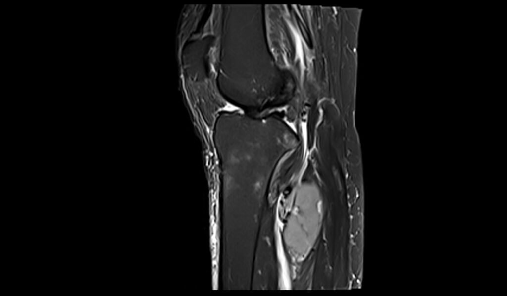
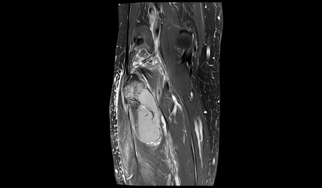
On Short Tau Inversion Recovery (STIR) sequences, knee lymphoma typically presents as hyperintense (bright) areas within the affected tissues. STIR sequences are highly sensitive to fluid and edema, allowing for clear visualization of the increased cellularity and associated edema within the lymphoma. The hyperintensity observed on STIR images helps in differentiating lymphoma from other types of soft tissue masses, as it highlights the extent of the tumor and any surrounding inflammatory response. This sequence is particularly useful for detecting small lesions and assessing the involvement of adjacent structures such as muscles and tendons.
MRI T1 Appearance of Knee Lymphoma
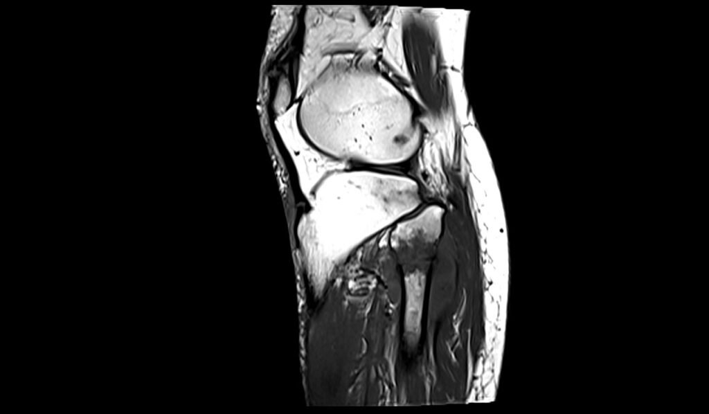
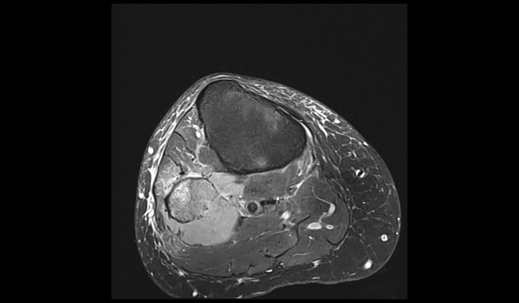
On T1-weighted MRI sequences, knee lymphoma generally appears as a hypointense (dark) mass compared to the surrounding muscle tissue. T1 sequences provide excellent anatomical detail, which helps in identifying the precise location and extent of the lymphoma. The hypointensity is due to the dense cellularity of the tumor, which differs from the intermediate signal intensity of normal muscle tissue. Post-contrast T1 images often show heterogeneous enhancement, reflecting the vascular nature of the tumor. This enhancement pattern can assist in differentiating lymphoma from other benign and malignant lesions.
MRI PD FS Appearance of Knee Lymphoma
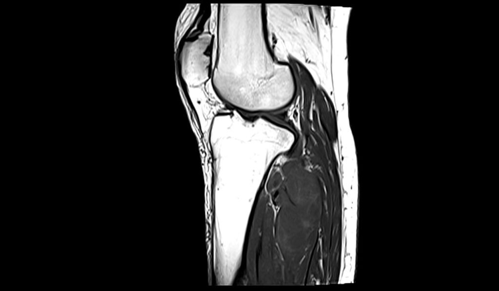
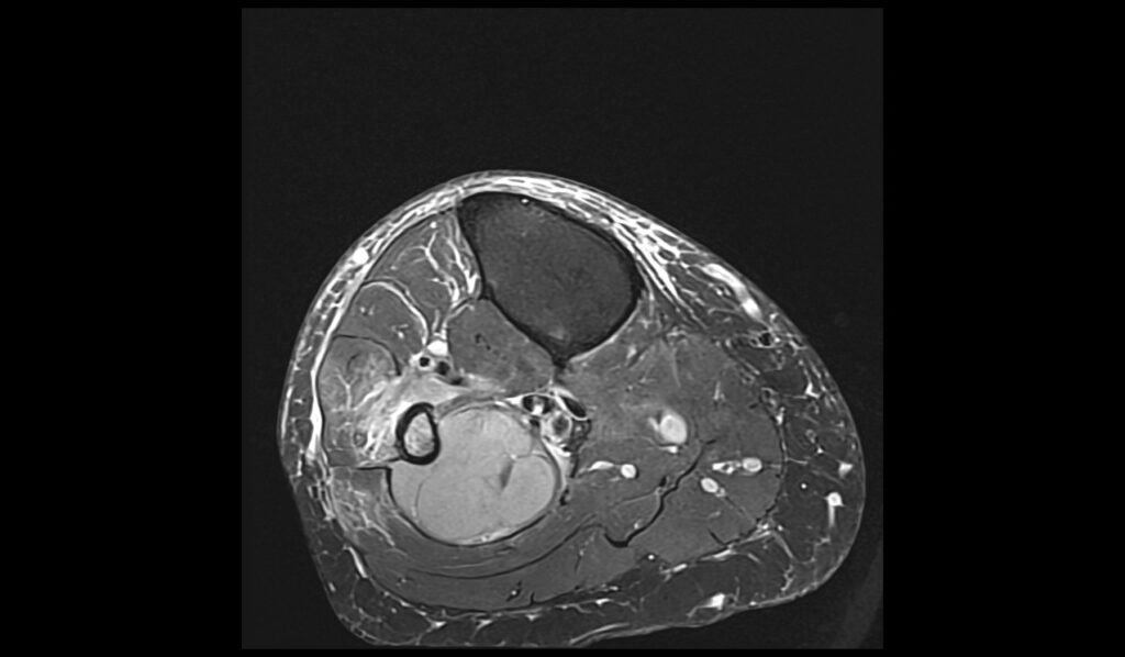
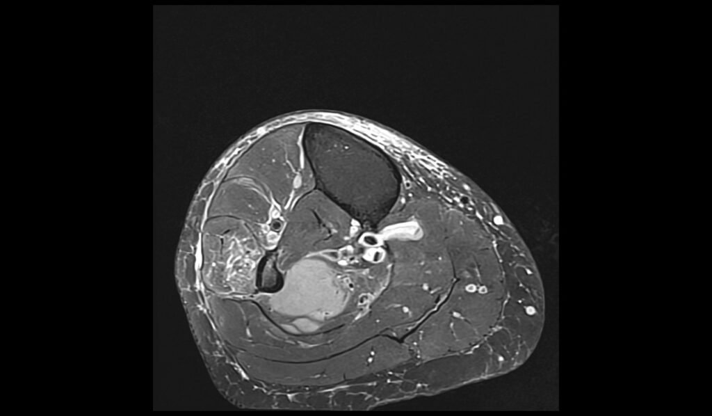
Proton Density Fat-Suppressed (PD FS) sequences are useful for visualizing knee lymphoma due to their ability to suppress the signal from fat and enhance the contrast of soft tissues. On PD FS images, lymphoma typically appears as an area of intermediate to hyperintense signal relative to the suppressed background. This sequence helps in delineating the extent of the tumor and assessing its impact on surrounding structures, such as the joint capsule, ligaments, and bone marrow. The fat suppression technique enhances the visibility of the lesion against the background, making it easier to detect subtle abnormalities.
STIR sagittal image shows Knee Lymphoma


T1 sagittal image shows Knee Lymphoma
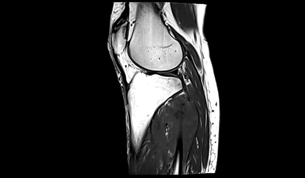
STIR coronal image shows Knee Lymphoma


PD FS axial image shows Knee Lymphoma

References
- Lim, C.Y., & Ong, K.O. (2013). Imaging of musculoskeletal lymphoma. Cancer Imaging, 13(4), 448-457. https://doi.org/10.1102/1470-7330.2013.0036.
- Wright, B., Moore, A., Becker-Weidman, D., & Joyce, D. (2024). Primary B cell lymphoma of the patella presenting as anterior knee pain: A case report. Journal of Orthopaedics, 48, 60-63. https://doi.org/10.1016/j.jor.2023.11.022
- Chun, K. A., Kim, H. J., Lee, S. H., Lee, S. H., Suh, J. S., Lee, J. H., … & Jee, W. H. (2010). Musculoskeletal Imaging: Original Research. AJR American Journal of Roentgenology, 195(6), 1355–1360.