MRI of Knee fracture
Knee fractures are serious injuries that occur when one of the bones in or around the knee joint breaks. These types of fractures can vary greatly in terms of severity, from small cracks in the bone to complete breaks that pierce the skin. The knee is a complex joint where the femur (thigh bone), tibia (shin bone), and patella (knee cap) meet, and a fracture can occur in any of these bones.
Causes of Knee Fractures
Knee fractures are typically the result of high-impact trauma. Common causes include:
- Vehicle Accidents: Collisions can exert extreme forces on the knee, causing fractures.
- Falls: High falls or slipping in an awkward position can result in a direct blow to the knee or an abnormal twisting motion leading to a fracture.
- Sports Injuries: Contact sports or activities involving jumping and running can lead to fractures due to direct impact or twisting forces on the knee joint.
- Osteoporosis: This condition weakens bones, making them more susceptible to fractures even with minor trauma.
- Overuse: Stress fractures can develop over time due to repeated strain or overuse, particularly in athletes.
Symptoms of Knee Fractures:
The symptoms of a knee fracture can vary depending on the severity and the specific bone involved. However, common signs include:
- Immediate and severe pain in the knee.
- Swelling and bruising around the knee.
- Inability to bear weight on the affected leg.
- Visible deformity or misalignment of the knee joint.
- Restricted range of motion in the knee.
- A feeling of instability or the knee giving way.
MRI Appearance of Knee fractures
T1-Weighted Images: On T1-weighted sequences, normal bone marrow is high in signal because of its fat content, and it appears bright. A fracture line may appear as a low signal intensity line (dark line) because the normal marrow fat is disrupted. Adjacent soft tissue swelling or hematoma also may be more apparent because of the contrast with the high signal fat.
Proton Density (PD) with Fat Saturation: PD imaging with fat saturation will suppress the high signal from fat, making fat appear dark. Edema, including the fluid associated with a fracture, will appear bright. This makes it easier to detect bone bruises or subtle fractures, as the edema associated with these injuries will stand out against the suppressed signal of the surrounding fat.
Short Tau Inversion Recovery (STIR): STIR images are very sensitive to fluid. This sequence inverts the signal from fat, making it appear dark, and any water or fluid in the tissue (such as from edema or hemorrhage) will appear bright. Fracture lines and bone bruises associated with the fracture are usually well-delineated due to the high water content in the traumatized area.
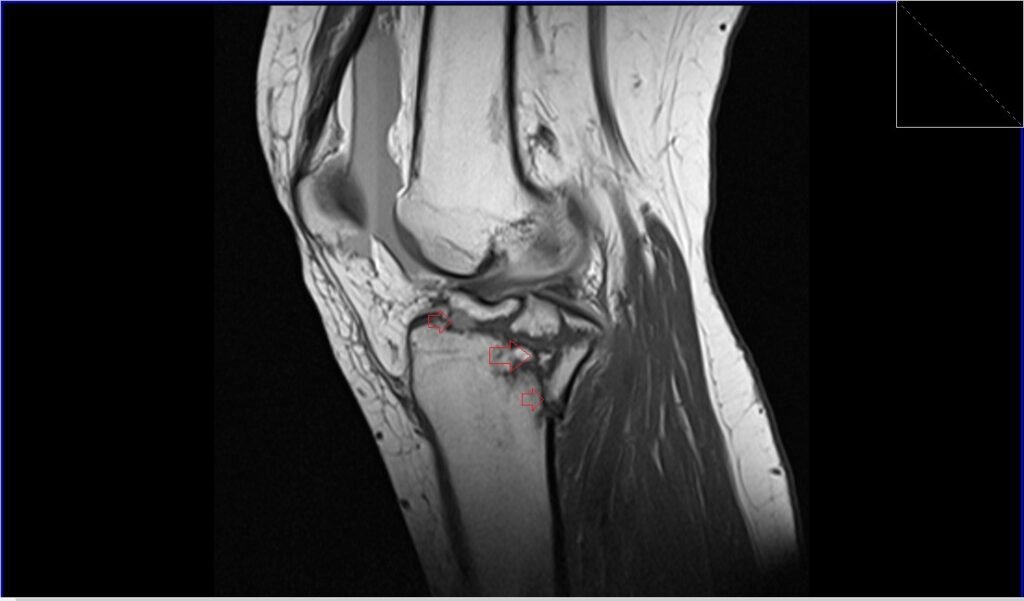
T1 TSE sagittal image shows knee fracture
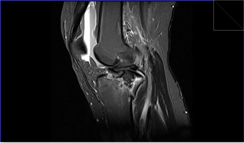
STIR sagittal image shows knee fracture
PD fat saturated coronal shows knee fracture
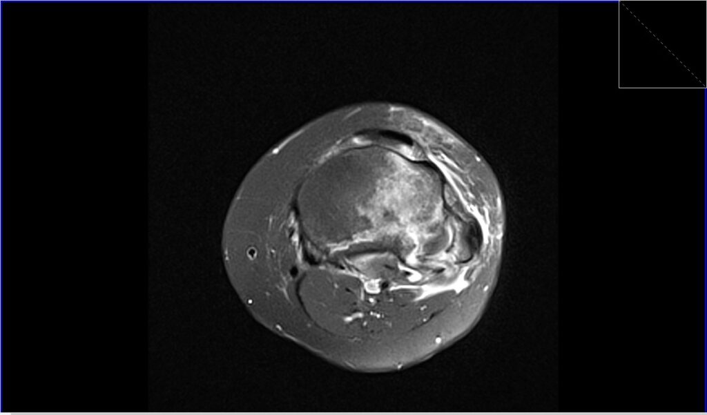
PD fat saturated axial image shows knee fracture
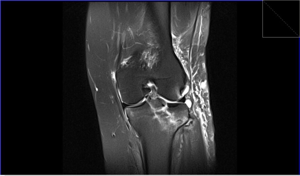
T1 TSE coronal image shows knee fracture

References
- Jarraya, M., Diaz, L. E., Arndt, W. F., Roemer, F. W., & Guermazi, A. (2017). Imaging of patellar fractures. Insights Imaging, 8(1), 49–57. doi: 10.1007/s13244-016-0535-0
- MacMahon, Peter J., and William E. Palmer. “A Biomechanical Approach to MRI of Acute Knee Injuries.” American Journal of Roentgenology, vol. 197, no. 3, September 2011
- Speer, K. P., Spritzer, C. E., & Garrett, W. E. Jr. (1991). Magnetic resonance imaging of traumatic knee articular cartilage injuries. The American Journal of Sports Medicine, 19(4)
- Oei, E.H.G., Ginai, A.Z., & Hunink, M.G.M. (2007). MRI for Traumatic Knee Injury: A Review. Seminars in Ultrasound, CT and MRI, 28(2), 141-157.