Infectious Myositis MRI
Infectious myositis is a condition characterized by inflammation of the muscles due to an infection. This condition can affect individuals of all ages and can be caused by various types of infectious agents, including bacteria, viruses, fungi, and parasites.
Causes
The primary causes of infectious myositis include:
- Bacteria: Common bacterial causes include Staphylococcus aureus, Streptococcus species, and Clostridium species.
- Viruses: Viral infections such as the influenza virus, HIV, and Coxsackievirus can lead to myositis.
- Fungi: Fungal infections, though less common, can also cause myositis, particularly in immunocompromised individuals.
- Parasites: Parasitic infections like Trichinella spiralis (which causes trichinosis) and Toxoplasma gondii can result in muscle inflammation.
Symptoms
The symptoms of infectious myositis can vary depending on the causative agent and the severity of the infection. Common symptoms include:
- Muscle pain and tenderness
- Swelling of the affected muscles
- Weakness in the muscles
- Fever
- Redness and warmth over the affected area
- Fatigue and general malaise
- In severe cases, abscess formation within the muscle tissue
Diagnosis
Diagnosis of infectious myositis typically involves a combination of clinical evaluation, laboratory tests, and imaging studies:
- Clinical Evaluation: Physical examination to assess muscle tenderness, swelling, and weakness.
- Laboratory Tests: Blood tests to check for elevated levels of muscle enzymes (such as creatine kinase), white blood cell count, and markers of inflammation. Blood cultures may be performed to identify the causative organism.
- Imaging Studies: MRI or CT scans can help visualize muscle inflammation and identify any abscesses.
- Muscle Biopsy: In some cases, a biopsy of the affected muscle may be necessary to identify the infectious agent and confirm the diagnosis.
Treatment
Treatment for infectious myositis depends on the underlying cause and the severity of the condition:
- Antibiotics: For bacterial infections, appropriate antibiotics are prescribed based on the identified pathogen and its antibiotic sensitivity.
- Antiviral Medications: In cases of viral myositis, antiviral medications may be used, especially in severe cases or those caused by specific viruses like HIV.
- Antifungal Medications: Fungal infections are treated with antifungal medications.
- Antiparasitic Medications: Parasitic myositis is treated with antiparasitic drugs.
- Surgical Intervention: In cases where abscesses have formed, surgical drainage may be necessary.
MRI Appearance of Infectious Myositis
MRI STIR Appearance of Infectious Myositis
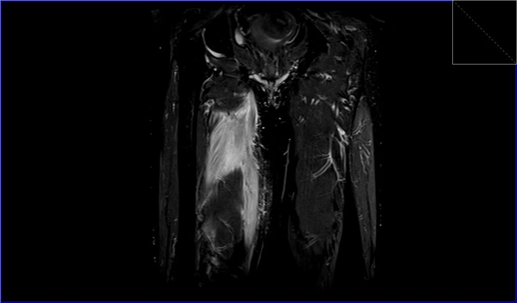
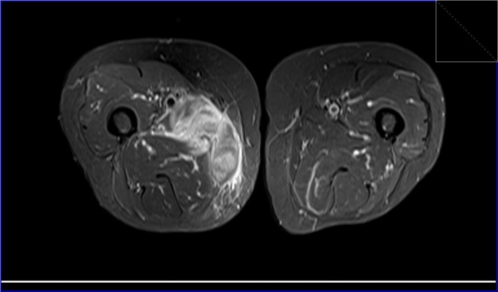
On STIR (Short Tau Inversion Recovery) sequences, infectious myositis typically presents with high signal intensity within the affected muscles due to the presence of edema and inflammation. The increased signal intensity is a result of the high water content associated with the inflammatory process, making STIR sequences particularly sensitive to detecting these changes. This hyperintensity helps to delineate the extent of the infection and inflammation within the muscle tissue, providing crucial information for the diagnosis and assessment of infectious myositis.
MRI T1 Appearance of Infectious Myositis
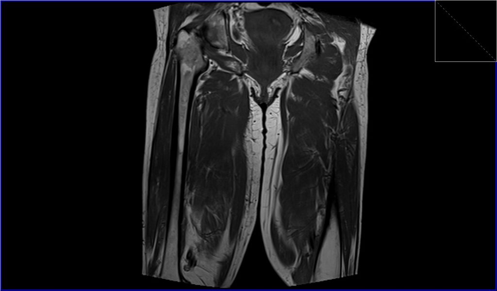
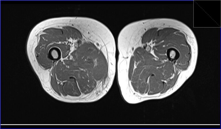
Infectious myositis on T1-weighted MRI images generally appears as areas of low to intermediate signal intensity within the affected muscles compared to normal muscle tissue. T1-weighted images are less sensitive to changes in water content, so the muscle tissue in the inflamed region typically appears darker than the surrounding healthy muscle. This imaging characteristic helps to identify the presence of muscle involvement and can be useful in assessing the overall structure and anatomy of the affected area, although it is less specific for inflammation and edema.
MRI T2 Appearance of Infectious Myositis
T2-weighted MRI images of infectious myositis show high signal intensity in the involved muscles due to the increased water content from edema and inflammatory exudate. This hyperintensity on T2 images is indicative of the inflammatory response and provides a clear contrast with the normal muscle tissue, which appears darker. T2-weighted sequences are effective in highlighting the extent of inflammation and fluid accumulation within the muscles, making them valuable in the evaluation and diagnosis of infectious myositis.
STIR coronal image of thigh shows Infectious Myositis
T1 TSE coronal image of thigh shows Infectious Myositis
STIR axial image of thigh shows Infectious Myositis
T1 TSE axial image of thigh shows Infectious Myositis
References
- Wasserman, P. L., Way, A., Baig, S., & Gopireddy, D. R. (2021). MRI of myositis and other urgent muscle-related disorders. Emerg Radiol, 28(2), 409–421. https://doi.org/10.1007/s10140-020-01866-2
- Schulze, M., Kötter, I., Ernemann, U., Fenchel, M., Tzaribatchev, N., Claussen, C. D., & Horger, M. (2009). MRI findings in inflammatory muscle diseases and their noninflammatory mimics. American Journal of Roentgenology, 192(6). https://doi.org/10.2214/AJR.08.1764
- Narayanappa, G., & Nandeesh, B. N. (2021). Infective myositis. 31(3), e12950. https://doi.org/10.1111/bpa.12950
- Martin, A., Aftab, S., Grewal, U., & Tom. (2015). Infectious myositis of the iliacus muscle: An important differential in the unwell child with a limp. J Case Rep Images Surg, 1, 17–20