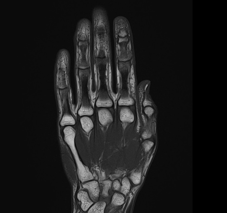
T1 MRI Sequence (T1 SE and TSE)
T1 (or longitudinal relaxation time) is a fundamental concept in MRI physics that refers to the time it takes for the longitudinal magnetization of a tissue to recover to its equilibrium state after being perturbed by an external radiofrequency (RF) pulse. T1 relaxation is a crucial parameter in MRI because it influences the contrast between different tissues in an MRI image.
Here’s a brief overview of the physics behind T1 relaxation in MRI:
Equilibrium Magnetization: In a magnetic field (B0), the protons (hydrogen nuclei) in a tissue align with the magnetic field. This creates a net macroscopic magnetization along the direction of the magnetic field, referred to as the equilibrium magnetization or the longitudinal magnetization (Mz).
Perturbation by RF Pulse: To create an MRI image, RF pulses are applied perpendicular to the B0 magnetic field. These pulses can tip the longitudinal magnetization away from the B0 direction, effectively moving it to the transverse plane (xy plane).
Relaxation Processes: After the RF pulse is turned off, the perturbed magnetization starts to recover back to its equilibrium state along the B0 direction. This recovery involves two main relaxation processes: T1 relaxation and T2 relaxation.
T1 Relaxation: T1 relaxation, also called longitudinal relaxation, is the recovery of the longitudinal magnetization (Mz) back towards its equilibrium value. This process is governed by the time constant T1. As the tissue’s protons return to their equilibrium alignment with the magnetic field, the longitudinal magnetization increases. Different tissues have different T1 values, leading to varying rates of recovery and influencing the contrast in MRI images.
T2 Relaxation: T2 relaxation, also called transverse relaxation, is the decay of the transverse magnetization (Mxy) due to interactions between neighboring protons. It is governed by the time constant T2. T2 relaxation leads to the loss of phase coherence among protons, resulting in signal decay in the transverse plane.
T1 Weighted and T2 Weighted Images: By manipulating the timing of RF pulses and the time between pulse sequences, MRI scanners can generate T1-weighted and T2-weighted images. T1-weighted images provide good anatomical detail, while T2-weighted images are sensitive to changes in tissue water content and can highlight pathologies like inflammation and edema.
MRI T1 image characteristics
When an MRI sequence is configured to generate a T1-weighted image, the tissues with short T1 values exhibit the highest magnetization and appear the brightest in the resulting image. A T1-weighted sequence enhances T1 contrast primarily by reducing the influence of T2 contributions. This is typically achieved by employing short repetition times (TR) of 300-600 ms to amplify the distinction in longitudinal relaxation during the return to equilibrium, and utilizing a short echo time (TE) of 10-15 ms to minimize the impact of T2 dependency during signal acquisition.
MRI T1 image appearance
The easiest way to identify T1-weighted images is to look for fluid-filled spaces in the body, such as cerebrospinal fluid in the brain ventricles and spinal canal, free fluid in the abdomen, fluid in the gall bladder and common bile duct, synovial fluid in joints, fluid in the urinary tract and urinary bladder, edema, or any other pathological fluid collection in the body. Fluids normally appear dark in a T1 weighted image. Here’s how different organs and tissues appear on T1-weighted images:
Fat-Rich Tissues:
- Fat-rich tissues, such as subcutaneous fat and bone marrow, appear bright or white on T1-weighted images due to their short T1 relaxation times. Fat suppresses the signal from surrounding tissues.
Muscles and Soft Tissues:
- Muscles and most soft tissues appear with intermediate signal intensity on T1-weighted images, often appearing as shades of gray.
Gray and White Matter in the Brain:
- In the brain, gray matter tends to appear slightly darker than white matter on T1-weighted images, reflecting differences in relaxation times and proton density.
Blood:
- Blood within vessels appears dark on T1-weighted images due to its long T1 relaxation time.
Fluid-Containing Structures:
- Fluid-filled structures, such as cysts, may appear dark or black on T1-weighted images due to their long T1 relaxation times.
Bone and Calcifications:
- Bone typically appears dark on T1-weighted images due to its very low proton density, minimal free water content, and extremely short T2 relaxation time. Calcifications also usually appear dark.
Liver:
- The liver often appears with intermediate signal intensity on T1-weighted images. It may show slight variations in signal intensity depending on its composition and fat content.
Spleen:
- The spleen typically exhibits similar signal intensity to the liver on T1-weighted images, reflecting its similar tissue composition.
Kidneys:
- The renal cortex (outer region of the kidneys) appears slightly brighter than the renal medulla (inner region) on T1-weighted images due to differences in tissue composition and water content.
Pancreas:
- The pancreas usually shows intermediate signal intensity on T1-weighted images, allowing for visualization of its structure and potential abnormalities.Bone typically appears dark on T1-weighted images due to its very low proton density, minimal free water content, and extremely short T2 relaxation time. Calcifications also usually appear dark.
Pathological appearance in T1 Weighted MRI
Pathological processes normally increase the water content in tissues. Due to the added water component, this results in a signal loss on T1-weighted images and a signal increase on T2-weighted images. Consequently, pathological processes are usually bright on T2-weighted images and dark on T1-weighted images.
The appearance of pathology on T1-weighted images can vary based on the specific tissue or abnormality. Here are several ways in which different pathologies could manifest on T1-weighted images:
Tumors: Tumors might exhibit altered signal intensity regions on T1-weighted images. Depending on factors such as the tumor’s cellularity, vascularity, and composition, they can appear either hypointense (darker) or isointense(grayish) in comparison to the surrounding tissues.
Edema: Edematous tissues could display hypointensity on T1-weighted images due to heightened water content. This is attributed to the longer T1 relaxation time of water, resulting in a reduction of signal intensity.
Hemorrhage: Hemorrhages can display different signal intensities at various stages:
- Hyperacute: Appears isointense.
- Acute: Transitions from isointense to hypointense.
- Early subacute: Progressively turns hyperintense.
- Late subacute: Consistently shows as hyperintense.
- Chronic:
- At the periphery, T1 is hypointense.
- At the centre, T1 is isointense.
lipomas: Lipomas consist of adipose tissue, which typically appears hyperintense on T1-weighted images due to its substantial fat content. This becomes especially noteworthy during imaging sequences that involve fat saturation techniques, causing the adipose tissue to appear darker.
Calcifications: Calcifications commonly emerge as hypointense on T1-weighted images. However, the specific appearance can differ based on the type and quantity of calcification present.
Cysts: Simple fluid-filled cysts might display hypointensity(dark) on T1-weighted images due to their elevated water content. Conversely, complex or hemorrhagic cysts could exhibit varying signal intensities.
Inflammation: Inflammatory tissues may appear hypointensity on T1-weighted images owing to heightened water content and changes within the tissue.
Degenerative Changes: Degenerated tissues, such as degenerative discs in the spine, could manifest as hypointense on T1-weighted images due to alterations in water content and tissue composition.
Tissues and their T1 appearance
Tissues and their T1 appearance
Brain:
- CSF: Dark.
- Fat: Bright.
- White Matter: Intermediate to bright.
- Gray Matter: Intermediate to dark.
- Bone (skull): Dark.
- Bone Marrow: Intermediate to bright.
- Blood Vessels: Mostly Dark; Depending on flow characteristics, can be bright or dark
- Pituitary Gland: Intermediate.
- Choroid Plexus: Intermediate.
- Cerebellum: Gray matter darker than white matter.
- Brain Stem: Intermediate to bright.
- Sinuses: Dark (air-filled).
- Thalamus, Putamen, Hippocampus, Caudate Nucleus: Intermediate.
- Corpus Callosum: Intermediate.
- Pineal Gland: Intermediate.
Spine:
- Spinal Cord: Intermediate.
- CSF: Dark.
- Bone: Dark.
- Bone Marrow: Intermediate to bright.
- Intervertebral Disc: Intermediate to bright
- Ligaments: Intermediate to Dark.
- Nerve Roots: Intermediate.
Abdomen and Pelvis:
- Liver: Intermediate to dark.
- Gallbladder and Common Bile Duct: Dark.
- Spleen: Intermediate.
- Kidney: Intermediate.
- Ureters: Intermediate.
- Pancreas: Intermediate to dark.
- Urinary Bladder: Dark.
- Prostate: Intermediate.
- Uterus: Intermediate.
Musculoskeletal:
- Muscle: Intermediate.
- Bone: Dark (low signal)
- Bone Marrow: Intermediate to bright
- Blood Vessels: Mostly Dark; Depending on flow characteristics, can be bright or dark
- Fat: Bright.
- Ligaments: Intermediate to dark
- Nerve Roots: Intermediate
- Cartilage: Intermediate to dark
- Synovial Fluid: Dark.
- Tendons: Intermediate to dark
Use
- Useful for abdominal imaging (T1 tse respiratory gated scans)
- Useful for chest imaging (T1 tse respiratory gated scans)
- Very useful for brachial and lumbar plexus imaging
- Very useful for anterior neck orbits and face imaging
- Very useful for any musculoskeletal imaging
- Very useful for extremity imaging
- Very useful for brain imaging
- Very useful for spine imaging
- Useful for pelvic imaging
MRI T1 SE axial image of brain

t1-weighted spin-echo axial image of pediatric brain

T1-weighted turbo spin-echo coronal image of the brain

T1-weighted turbo spin-echo coronal image of the orbits

T1-weighted turbo spin-echo axial image of the face

T1-weighted turbo spin-echo axial image of the neck

T1-weighted turbo spin-echo sagittal image of the cervical spine

MRI T1-weighted turbo spin-echo axial image of the chest

T1-weighted turbo spin-echo coronal image of the sternum
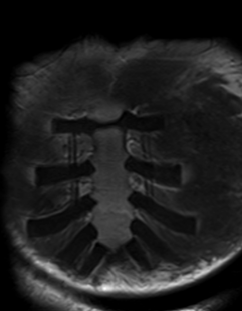
T1-weighted turbo spin-echo axial image of the thoracic spine

T1-weighted turbo spin-echo axial image of the lumbar spine

T1-weighted turbo spin-echo sagittal image of the lumbar spine

T1-weighted turbo spin-echo coronal image of the psoas muscle

T1-weighted turbo spin-echo axial image of the abdomen

T1-weighted turbo spin-echo axial image of the pelvis

T1-weighted turbo spin-echo coronal image of the SIJ

T1-weighted turbo spin-echo axial image of the penis

T1-weighted turbo spin-echo coronal image of the hips

T1-weighted turbo spin-echo coronal image of the thigh
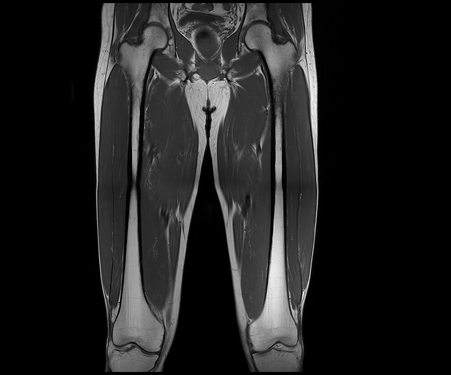
T1-weighted turbo spin-echo axial image of the thigh

T1-weighted turbo spin-echo sagittal image of the knee

T1-weighted turbo spin-echo axial image of the legs

T1-weighted turbo spin-echo sagittal image of the ankle

T1-weighted turbo spin-echo coronal image of the foot

T1-weighted turbo spin-echo axial image of the foot
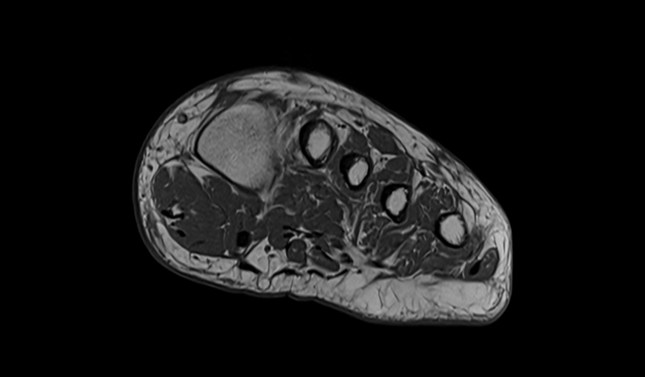
T1-weighted turbo spin-echo axial image of the shoulder

T1-weighted turbo spin-echo coronal image of the shoulder

T1-weighted turbo spin-echo coronal image of the upper arm
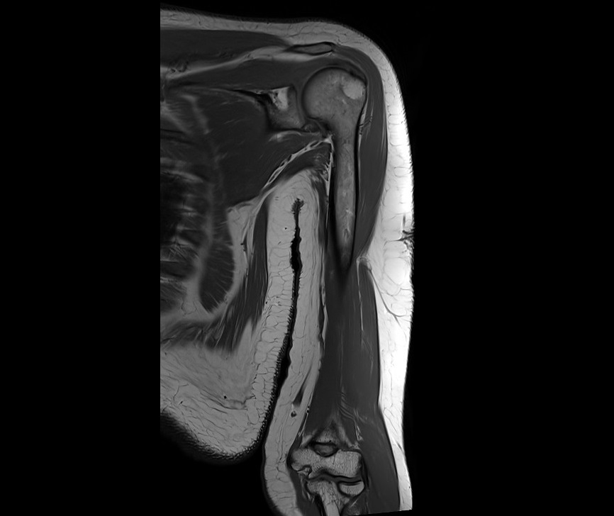
T1-weighted turbo spin-echo coronal image of the elbow

T1-weighted turbo spin-echo coronal image of the forearm
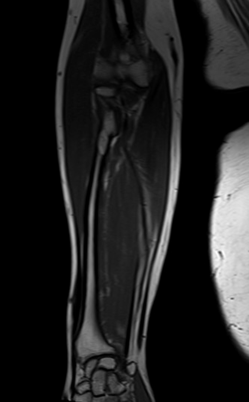
T1-weighted turbo spin-echo coronal image of the wrist

T1-weighted turbo spin-echo sagittal image of the hand
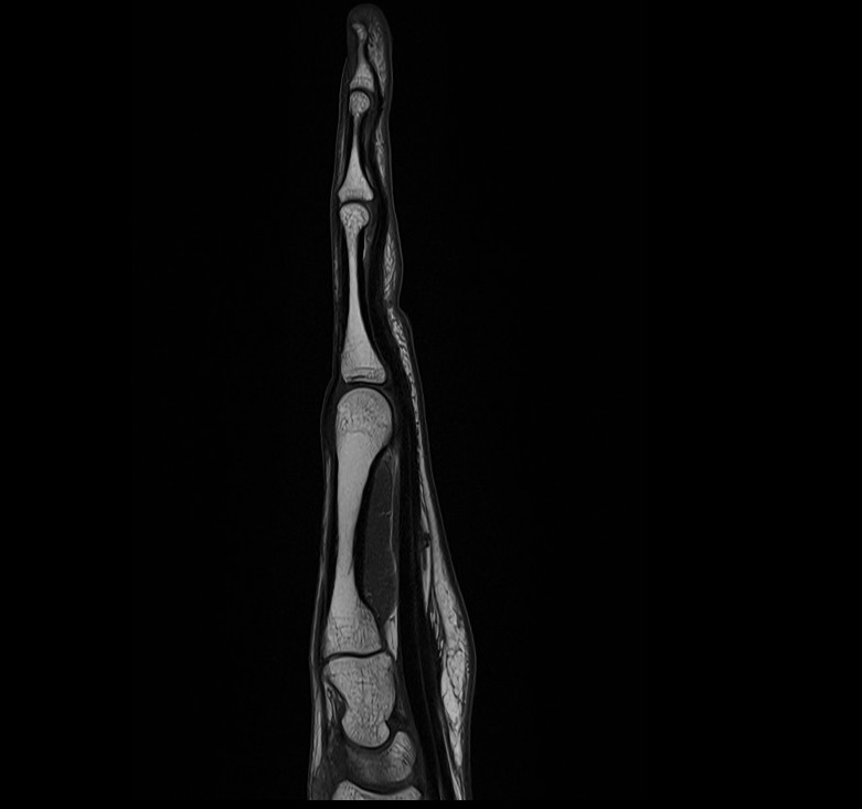
T1-weighted turbo spin-echo coronal image of the hand
