MRI Hepatocellular carcinoma (HCC)
Hepatocellular carcinoma (HCC) is the most common type of primary liver cancer, occurring predominantly in patients with underlying chronic liver disease and cirrhosis. It arises from hepatocytes, the liver’s main cell type, which makes up about 80% of the liver’s mass. Understanding HCC involves looking at its causes, symptoms, and available treatments.
Causes:
The development of HCC is typically linked to several risk factors:
- Chronic viral hepatitis: Hepatitis B and C are significant risk factors, with chronic infection leading to liver damage and increased cancer risk.
- Cirrhosis: Regardless of the cause, cirrhosis (scarring of the liver) enhances the risk of developing HCC.
- Alcohol consumption: Excessive alcohol intake can lead to alcoholic liver disease and cirrhosis, subsequently increasing HCC risk.
- Other Factors: These include hereditary diseases such as hemochromatosis, autoimmune hepatitis, and primary biliary cirrhosis, as well as lifestyle factors such as obesity and type 2 diabetes.
Symptoms:
HCC usually remains asymptomatic in its early stages and is often diagnosed incidentally during routine screening in patients with known risk factors. As the tumor grows, symptoms may include:
- Abdominal Pain: Particularly in the upper right part of the abdomen.
- Weight Loss: Unintentional and significant weight loss is common.
- Weakness and Fatigue: General feelings of being unwell.
- Jaundice: Yellowing of the skin and eyes due to liver dysfunction.
- Swelling: Accumulation of fluid in the abdomen (ascites) or swelling of the legs.
- Others: Loss of appetite, nausea, or vomiting.
Treatment:
The treatment of HCC depends on several factors, including the stage of the disease, liver function, overall health, and the presence of cirrhosis. Options include:
- Surgical Resection: Removal of the tumor is possible in patients with good liver function and a solitary tumor with no vascular invasion.
- Liver Transplantation: Suitable for patients with early-stage HCC who have significant liver dysfunction or cirrhosis.
- Targeted Therapy: Drugs such as sorafenib, lenvatinib, and others block the growth and spread of cancer by interfering with specific molecules involved in tumor growth and progression. These are generally used for advanced HCC.
- Immunotherapy: Treatments like nivolumab and pembrolizumab help the immune system recognize and attack cancer cells. These are options for patients who do not respond to targeted therapies.
- Radiation Therapy: Advanced techniques like stereotactic body radiation therapy (SBRT) can deliver high doses of radiation to the tumor with minimal impact on surrounding healthy tissues, used mainly when other treatments are not suitable.
- Chemotherapy: Less commonly used for HCC compared to other cancers, as it tends to be less effective. However, it may be used in certain circumstances
- Ablation therapies: Techniques like radiofrequency ablation or microwave ablation destroy the tumor without removing it.
MRI Hepatocellular carcinoma (HCC)
T1-Weighted Imaging
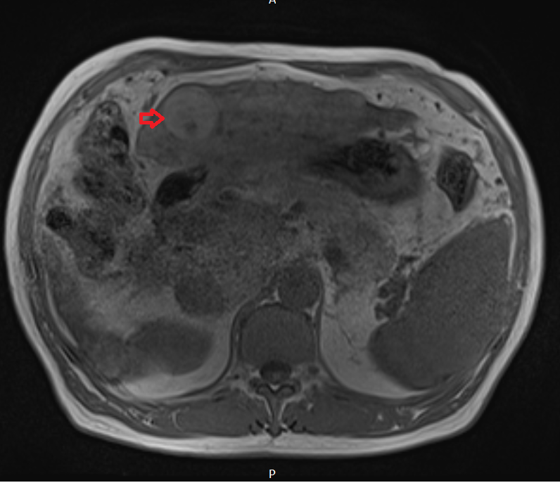
- T1-Weighted: HCC usually appears hypointense (darker) on T1-weighted images compared to the surrounding liver tissue, although some well-differentiated HCCs can be hyperintense (brighter) due to their high fat content.
In-Phase and Out-Phase Imaging
- In-Phase/Out-Phase: These sequences are useful in detecting fat in liver lesions. HCC can show signal loss on out-phase images compared to in-phase due to the presence of microscopic fat within the tumor.
T2-Weighted Imaging
- Low TE (Echo Time): T2-weighted images with a shorter TE can provide clearer details of the tumor structure. HCC usually appears slightly hyperintense on these images.
- High TE: With a longer TE, fluid characteristics become more pronounced, making HCC appear more prominently hyperintense compared to surrounding liver tissue.
STIR (Short TI Inversion Recovery)
- HCC lesions can appear hyperintense on STIR images. This sequence is particularly useful for suppressing the background signal from fat, which helps in differentiating HCC from surrounding liver tissue.
DWI (Diffusion Weighted Imaging)
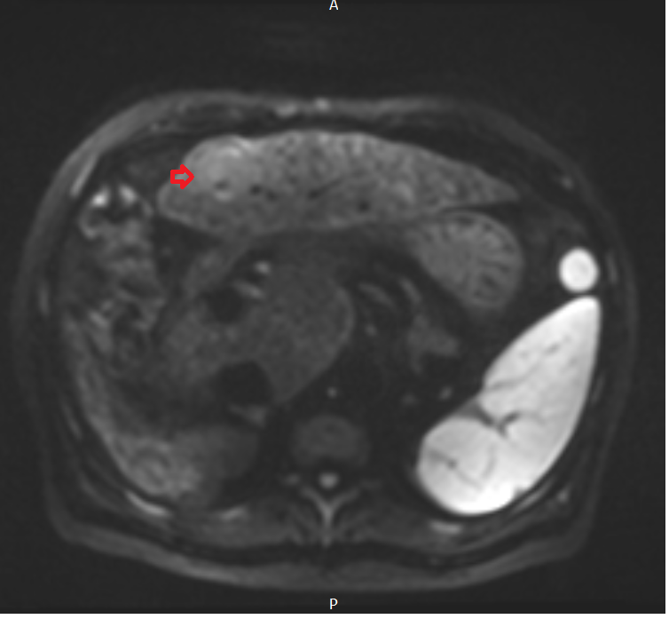
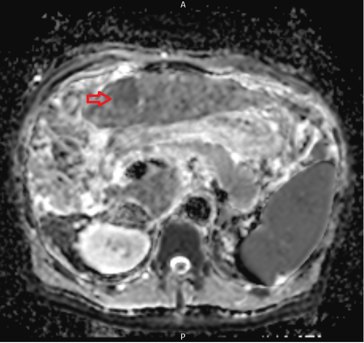
- HCC often shows high signal intensity on high b-value DWI images and low apparent diffusion coefficient (ADC) values, reflecting the restricted diffusion within malignant tissues.
Post-Contrast T1-Weighted Dynamic Imaging
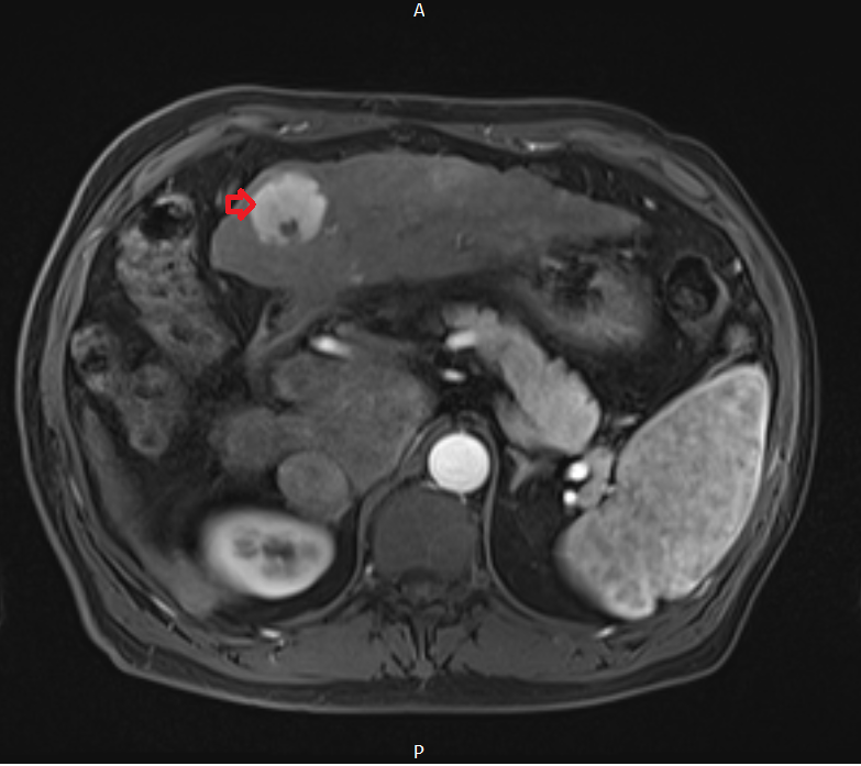
- Arterial Phase: HCC shows rapid enhancement due to its arterial blood supply.
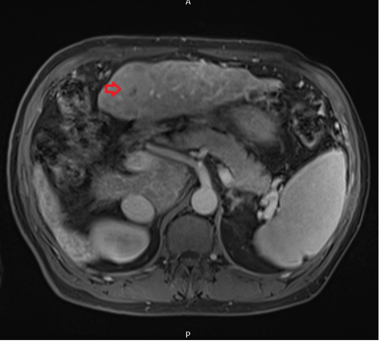
- Venous Phase: There’s typically a noticeable washout of contrast in HCC, which is less enhanced than the liver at this phase.
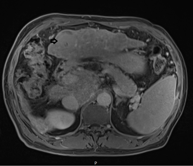
- Delayed Phase: Continued washout makes HCC appear hypointense compared to the surrounding liver parenchyma.
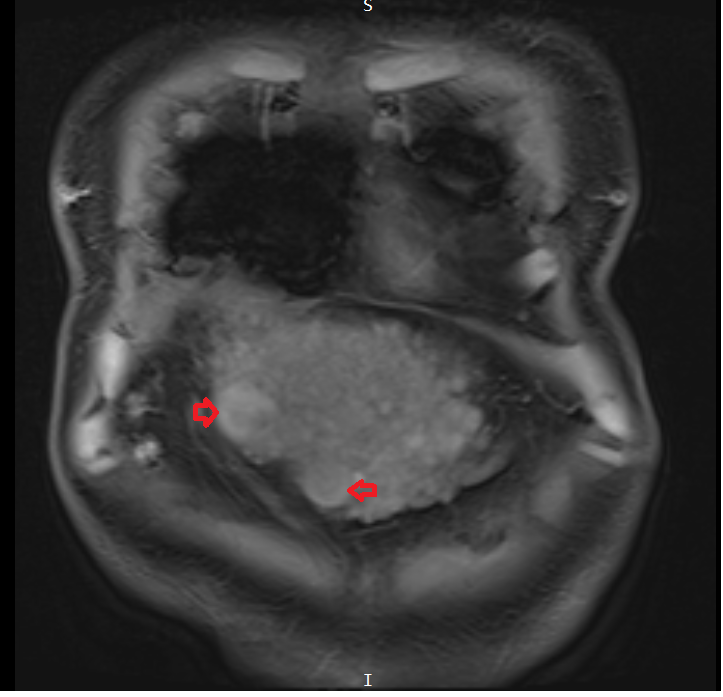
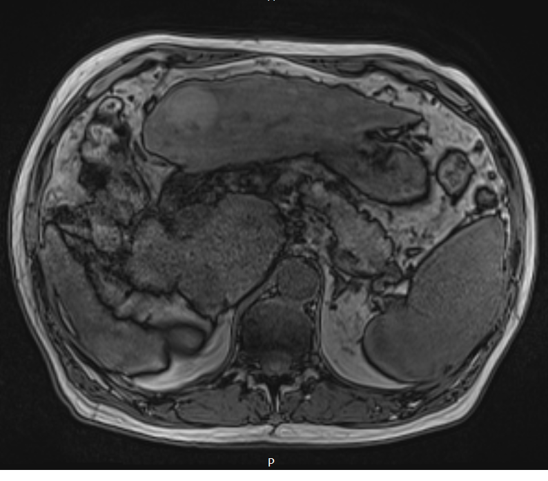
T1 VIBE coronal image shows Hepatocellular carcinoma (HCC)
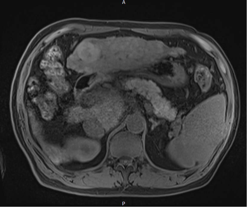
T1 VIBE axial image shows Hepatocellular carcinoma (HCC)
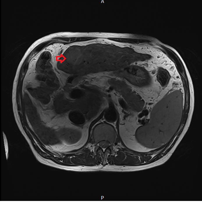
T2 TE 90 axial image shows Hepatocellular carcinoma (HCC)
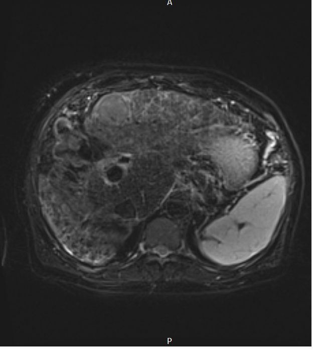
T2 TE 190 axial image shows Hepatocellular carcinoma (HCC)

T2 STIR axial image shows Hepatocellular carcinoma (HCC)
T1 in-phase axial image shows Hepatocellular carcinoma (HCC)
T1 out-of-phase axial image shows Hepatocellular carcinoma (HCC)
DWI b50 axial image shows Hepatocellular carcinoma (HCC)
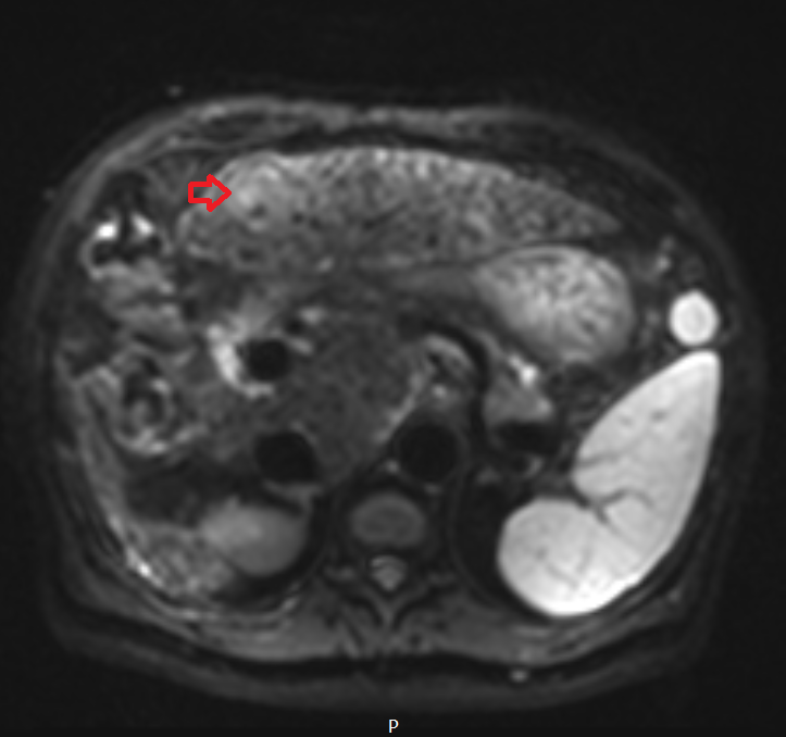
DWI b800 axial image shows Hepatocellular carcinoma (HCC)
DWI ADC axial image shows Hepatocellular carcinoma (HCC)
T1 VIBE axial dynamic arterial phase image shows Hepatocellular carcinoma (HCC)
T1 VIBE axial dynamic portal venous phase image shows Hepatocellular carcinoma (HCC)
T1 VIBE axial dynamicdelayed phase image shows Hepatocellular carcinoma (HCC)
References
- Mendiratta-Lala, M., Masch, W. R., Shampain, K., Zhang, A., Jo, A. S., Moorman, S., Aslam, A., Maturen, K. E., & Davenport, M. S. (2020). MRI Assessment of Hepatocellular Carcinoma after Local-Regional Therapy: A Comprehensive Review. Radiology: Imaging Cancer, 2(1). Retrieved from RSNA website: https://doi.org/10.1148/rycan.2020190024
- Criss, C., Nagar, A. M., & Makary, M. S. (2023). Hepatocellular carcinoma: State of the art diagnostic imaging. World Journal of Radiology, 15(3), 56–68. https://doi.org/10.4329/wjr.v15.i3.5
- Coenegrachts, K. (2009). Magnetic resonance imaging of the liver: New imaging strategies for evaluating focal liver lesions. World Journal of Radiology, 1(1), 72–85. https://doi.org/10.4329/wjr.v1.i1.72
- chikawa, S., & Goshima, S. (2023). Clinical Significance of Liver MR Imaging. Magn Reson Med Sci, 22(2), 157–175. https://doi.org/10.2463/mrms.rev.2022-0100
- Tani, I., Kurihara, Y., Kawaguchi, A., Nakajima, Y., Ishikawa, T., Maeyama, S., & Tanaka, R. (2000). MR Imaging of Diffuse Liver Disease. American Journal of Roentgenology, 174(4). Retrieved from https://doi.org/10.2214/ajr.174.4.1740965