Hand Arteriovenous Malformations (AVMs) MRI
Arteriovenous malformations (AVMs) are abnormal connections between arteries and veins, bypassing the capillary system. In the hand, these malformations can be particularly troublesome due to the complexity and density of blood vessels in this area.
Causes
The exact cause of AVMs is not fully understood, but they are thought to occur due to developmental abnormalities during embryonic growth. Some AVMs may be present at birth (congenital), while others may develop later in life, potentially triggered by trauma or other unknown factors.
Symptoms
The symptoms of hand AVMs can vary widely depending on the size and location of the malformation. Common symptoms include:
- Pain: Often the most common symptom, which can range from mild to severe.
- Swelling: The hand may appear swollen, especially when the AVM is large.
- Pulsation: A throbbing or pulsating sensation in the affected area.
- Skin Changes: The skin over the AVM may appear reddish or purplish and warmer than the surrounding skin.
- Ulceration: In severe cases, the skin over the AVM may break down, leading to sores or ulcers.
- Functional Impairment: Depending on the size and location, the AVM can interfere with normal hand function, leading to weakness or difficulty in movement.
Diagnosis
Diagnosing AVMs typically involves a combination of physical examination and imaging studies. Diagnostic methods include:
- Ultrasound: To assess blood flow and the structure of the AVM.
- Magnetic Resonance Imaging (MRI): Provides detailed images of blood vessels and surrounding tissues.
- Computed Tomography (CT) Scan: Offers detailed cross-sectional images and can be used with angiography to visualize blood vessels.
- Angiography: Considered the gold standard for detailed visualization of the vascular architecture of AVMs. This involves injecting a contrast dye into the bloodstream and taking X-rays to trace the flow of blood through the arteries and veins.
Treatment
Treatment of hand AVMs is guided by the symptoms, size, and location of the vascular malformation. Options include:
- Observation: Small, asymptomatic AVMs may only require regular monitoring.
- Medication: To manage symptoms like pain or to reduce the risk of complications.
- Sclerotherapy: Injection of a sclerosing agent directly into the AVM to reduce its size and blood flow.
- Embolization: A minimally invasive procedure where materials are injected to block the abnormal blood vessels, reducing blood flow to the AVM.
- Surgery: Considered for larger or symptomatic AVMs. This involves the physical removal of the malformation, which can be challenging due to the risk of significant bleeding and the need to preserve hand function.
- Laser Therapy: Sometimes used for smaller or superficial AVMs.
MRI Appearance of Hand Arteriovenous Malformations (AVMs)
STIR Appearance of Hand Arteriovenous Malformations (AVMs)
On STIR (Short Tau Inversion Recovery) MRI sequences, arteriovenous malformations (AVMs) in the hand typically present as hyperintense, serpiginous structures due to the high signal intensity of blood flow within the abnormal vascular channels. The STIR sequence is highly sensitive to fluid, helping to delineate the AVMs from surrounding tissues.
T1 Appearance of Hand Arteriovenous Malformations (AVMs)
On T1-weighted MRI sequences, hand AVMs usually appear as isointense or slightly hypointense compared to the surrounding musculature. The vessels themselves might not be as conspicuous on T1-weighted images unless there is a significant amount of slow-flowing blood or thrombosis within the malformation. The anatomical detail provided by T1 sequences is essential for understanding the extent of the lesion, its relationship to adjacent structures, and for assessing any associated tissue abnormalities or complications such as hemosiderin deposition due to recurrent hemorrhage.
T1 Post-Contrast Appearance of Hand AVM’s
Following the administration of gadolinium-based contrast agents, T1-weighted post-contrast images enhance the visualization of hand AVMs. These lesions typically show intense, heterogeneous enhancement due to the rapid filling of the abnormal vessels with contrast material. The post-contrast images highlight the vascular architecture of the AVM, including the nidus, feeding arteries, and draining veins, providing critical information for evaluating the extent and nature of the malformation. This enhancement pattern is crucial for differentiating AVMs from other vascular or soft tissue anomalies.
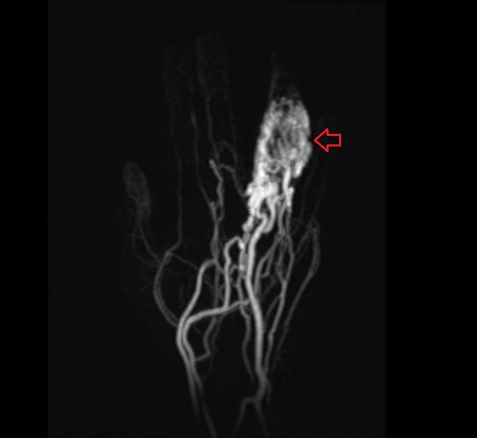
Dynamic MRA Appearance of Hand AVM’s
Dynamic Magnetic Resonance Angiography (MRA) offers detailed visualization of the vascular anatomy and hemodynamics of hand AVMs. This imaging modality captures the flow of contrast through the vascular system in real-time, revealing the rapid arterial filling followed by venous drainage characteristic of AVMs. The dynamic MRA sequences allow for the precise mapping of the feeding arteries, the nidus, and the draining veins, which is essential for interventional planning. The ability to visualize blood flow dynamics helps in assessing the shunt severity and planning targeted embolization or surgical interventions.
T1 coronal image of hand shows Arteriovenous Malformation (AVM)

T1 post contrast axial image of hand shows Arteriovenous Malformation (AVM)


T1 ppst contrast coronal dynamic image of hand shows Arteriovenous Malformation (AVM)


Post contrast coronal MIP image of hand shows Arteriovenous Malformation (AVM)
References
- Bashir, U., Shah, S., Jeph, S., O’Keeffe, M., & Khoso, F. (2017). Magnetic Resonance (MR) Imaging of Vascular Malformations. Polish Journal of Radiology, 82, 731–741. https://doi.org/10.12659/PJR.903491
- Sawani, A., Huber, K., Zibadi, S., & Payne, W. G. (2017). Diagnosis of Arteriovenous Malformation in the Finger. Eplasty, 17, ic10. https://www.ncbi.nlm.nih.gov/pmc/articles/PMC5408223/
- Drapé, J.-L., Feydy, A., Guerini, H., Godefroy, D., Le Viet, D., & Chevrot, A. (2005). Vascular lesions of the hand. European Journal of Radiology, 56(3), 331-343. https://doi.org/10.1016/j.ejrad.2005.03.013