Hamstring Injury MRI
A hamstring injury refers to the strain or tear of the hamstring muscles located at the back of the thigh. These muscles include the biceps femoris, semitendinosus, and semimembranosus. Hamstring injuries are common in athletes, especially those involved in sports that require sprinting, jumping, and sudden changes in direction.
Causes
- Overuse: Repeated stress on the hamstring muscles from activities like running, jumping, or cycling.
- Sudden Movements: Quick starts and stops or sudden changes in direction can strain the muscles.
- Muscle Imbalance: Weaker hamstrings compared to the quadriceps can lead to injury.
- Poor Flexibility: Tight muscles are more prone to strains.
- Inadequate Warm-Up: Not properly warming up before intense physical activity can increase the risk.
Symptoms
- Pain: Sudden and sharp pain in the back of the thigh during activity.
- Swelling: Swelling and bruising in the affected area.
- Weakness: Weakness in the hamstring muscles.
- Difficulty Walking: Pain and discomfort while walking or bending the knee.
- Tenderness: Sensitivity to touch in the injured area.
Diagnosis
- Physical Examination: A doctor will examine the injured area, assessing pain, swelling, and range of motion.
- Imaging Tests: MRI or ultrasound may be used to confirm the extent of the injury and rule out other possible conditions.
MRI Appearance of Hamstring Injury
MRI STIR Appearance of Hamstring Injury
The Short Tau Inversion Recovery (STIR) sequence in MRI is particularly sensitive for detecting edema and inflammation. In the context of a hamstring injury, the STIR sequence will typically show hyperintense (bright) signals in the region of the injured muscle, indicating the presence of edema or hemorrhage. This increased signal intensity helps in identifying the extent of the muscle damage, including partial or complete tears. The high contrast between the inflamed tissues and the surrounding normal muscles makes STIR an effective tool for early diagnosis and for assessing the severity of acute muscle injuries.
MRI T2 Appearance of Hamstring Injury
T2-weighted MRI images are highly sensitive to changes in water content and are excellent for visualizing acute muscle injuries. In the case of a hamstring injury, T2 images will show hyperintense (bright) signals in the affected area, indicating edema and inflammation. This sequence can also help in distinguishing between acute and chronic injuries, as acute injuries will present with brighter signals due to the presence of increased fluid. T2-weighted images are valuable for assessing the extent of the injury, including the involvement of tendons and the presence of any associated muscle tears or hematomas.
MRI PD FS Appearance of Hamstring Injury
Proton Density Fat-Suppressed (PD FS) MRI sequences combine the benefits of high-resolution anatomical detail with enhanced visualization of fluid and edema by suppressing the signal from fat. In a hamstring injury, PD FS images typically reveal hyperintense (bright) areas corresponding to the presence of edema and hemorrhage within the muscle, similar to T2-weighted images but with better contrast and detail. The fat suppression technique allows for a clearer view of the muscle tissue by eliminating the bright signal from fat, thereby highlighting the injured regions more effectively. This sequence is particularly useful for detecting subtle injuries and for assessing the integrity of muscle fibers and tendons within the hamstring group.
MRI T1 Appearance of Hamstring Injury
T1-weighted MRI images provide detailed anatomical information but are less sensitive to changes in water content compared to STIR and T2 sequences. In a hamstring injury, T1 images may show a disruption in the normal muscle architecture and may appear hypointense (darker) if there is significant hemorrhage or fibrosis. T1-weighted imaging is useful for visualizing the anatomy of the muscle and for detecting chronic changes such as fatty infiltration or scar tissue formation, which are indicative of a previous injury or a more prolonged healing process.
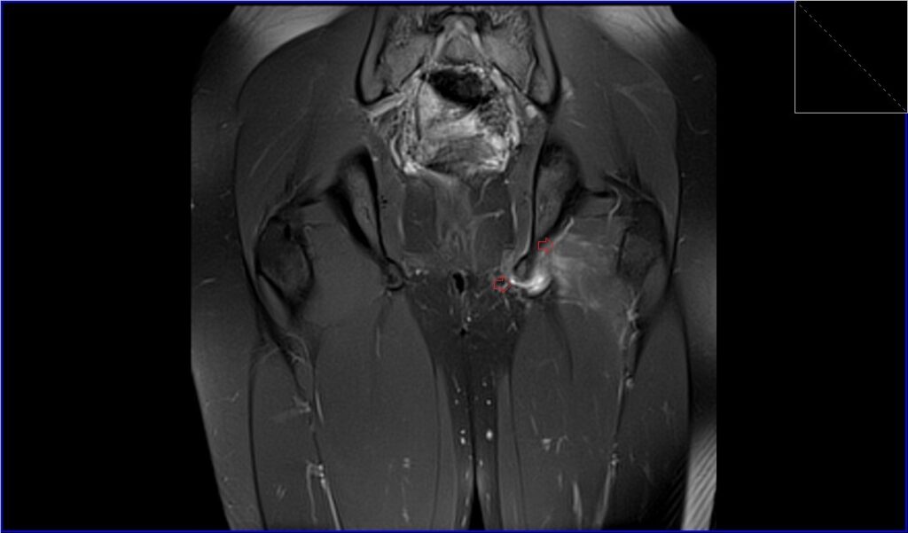
Proton Density Fat-Suppressed image of thigh shows Hamstring Injury
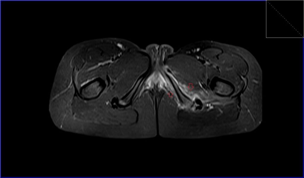
STIR axial image of thigh shows Hamstring Injury
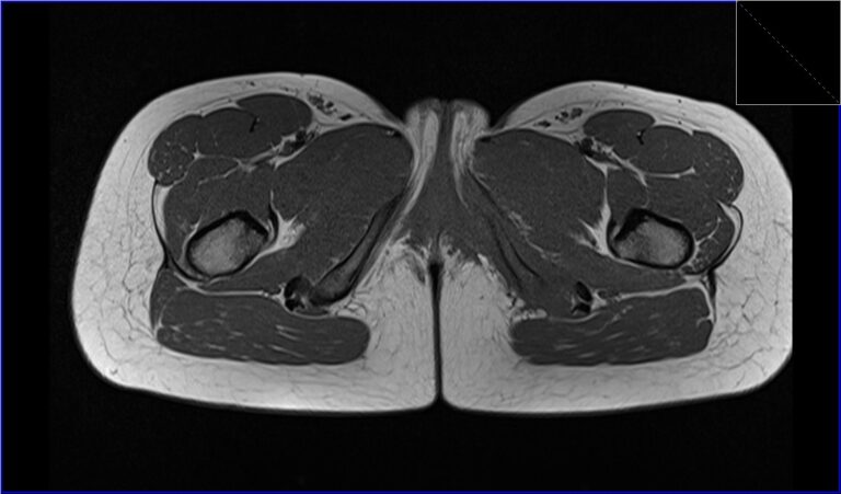
T1 TSE axial image of thigh shows Hamstring Injury
References
- Greenky, M., & Cohen, S. B. (2017). Magnetic resonance imaging for assessing hamstring injuries: Clinical benefits and pitfalls – a review of the current literature. Open Access Journal of Sports Medicine, 8, 167–170.
- Rubin, D. A. (2012). Imaging diagnosis and prognostication of hamstring injuries. American Journal of Roentgenology, 199(3). https://doi.org/10.2214/AJR.12.8784
- Pedret, C. (2021). Hamstring muscle injuries: MRI and ultrasound for diagnosis and prognosis. Journal of the Belgian Society of Radiology, 105(1), 91. https://doi.org/10.5334/jbsr.2617
- Gibbs, N. J., Cross, T. M., Cameron, M., & Houang, M. T. (2004). The accuracy of MRI in predicting recovery and recurrence of acute grade one hamstring muscle strains within the same season in Australian Rules football players. Journal of Science and Medicine in Sport, 7(2), 248-258.