MRI of Greater Tuberosity Avulsion Fracture
A greater tuberosity avulsion fracture is a type of shoulder injury where the greater tuberosity of the humerus is forcibly detached or fractured. This can occur due to direct trauma or indirectly through muscular contraction.
Causes
Greater tuberosity avulsion fractures are most commonly associated with:
- Direct trauma: Such as a fall on an outstretched arm or a direct blow to the shoulder.
- Indirect trauma: This can happen from sudden, forceful lifting or pulling movements, often during sports or strenuous activities.
Symptoms
The primary symptoms of a greater tuberosity avulsion fracture include:
- Pain: Especially when lifting the arm or during shoulder movements; the pain can be severe and is often localized at the top and outer side of the shoulder.
- Swelling and tenderness: Around the shoulder, particularly over the greater tuberosity.
- Reduced range of motion: Difficulty in raising the arm sideways or rotating it, often due to pain and mechanical blockage by the displaced bone fragment.
- Audible pop or snap: In some cases, there might be an audible sound at the time of injury.
- Weakness: In the shoulder, especially when trying to lift or rotate the arm.
Diagnosis
Diagnosis typically involves a combination of clinical examination and imaging studies:
- Physical examination: To assess pain, tenderness, range of motion, and strength in the shoulder.
- X-rays: Are the primary imaging tool used to confirm the presence of a fracture and to evaluate its location and severity.
- MRI: May be used if the involvement of soft tissue, such as rotator cuff tendons, is suspected or to assess the integrity of the surrounding structures.
Treatment
Treatment of greater tuberosity avulsion fractures depends on the size of the bone fragment and the degree of displacement:
- Non-surgical treatment: Includes rest, ice, and immobilization of the shoulder with a sling. Pain management often involves analgesics and anti-inflammatory medications. Physical therapy is crucial and begins with gentle range-of-motion exercises, gradually progressing to strengthening exercises once healing allows.
- Surgical treatment: May be required for large displacements or if the fracture disrupts shoulder function significantly. Surgery typically involves reattaching the bone fragment with screws or other fixation devices and repairing any associated rotator cuff injuries.
MRI Appearance of Greater Tuberosity Avulsion Fracture
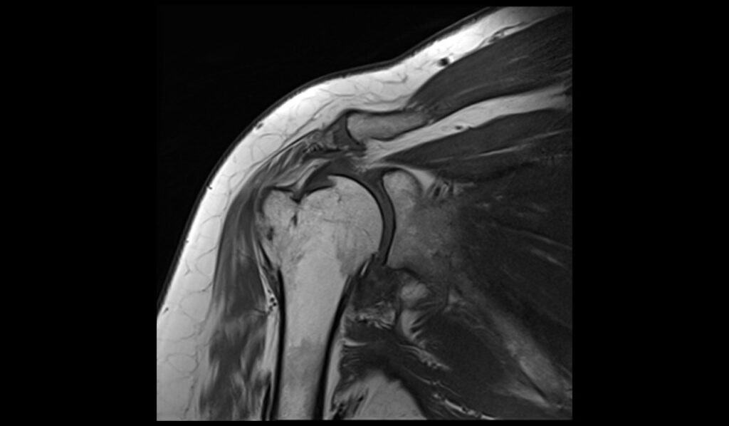
MRI T1 Appearance of Greater Tuberosity Avulsion Fracture
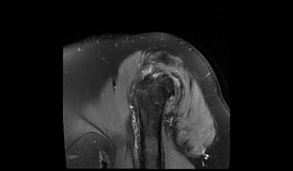
On T1-weighted MRI images, a greater tuberosity avulsion fracture typically appears as a hypointense (dark) line or zone at the site of the fracture. The surrounding bone marrow may show varying degrees of hypointensity due to hemorrhage or edema. In some cases, the fragment of the avulsed bone may be visible, displaced from its normal anatomical position. The intact cortical bone and the surrounding soft tissues, including the rotator cuff tendons, can also be assessed for any associated injuries or abnormalities.
MRI T2 Appearance of Greater Tuberosity Avulsion Fracture
T2-weighted MRI images are sensitive to fluid and inflammation, making them useful for identifying bone and soft tissue injuries. In the case of a greater tuberosity avulsion fracture, T2 images typically show a hyperintense (bright) signal at the fracture site due to the presence of bone marrow edema and hemorrhage. The surrounding soft tissues, such as the rotator cuff tendons, may also appear hyperintense if there is associated inflammation or injury. Additionally, fluid accumulation in the subacromial-subdeltoid bursa may be observed if there is significant soft tissue damage.
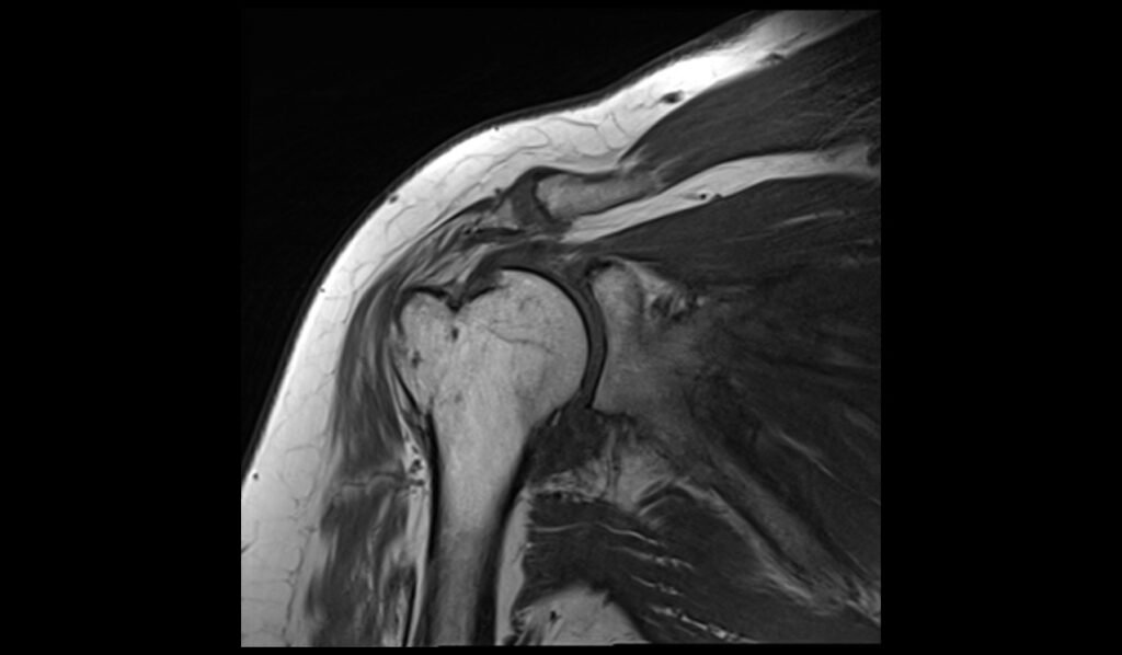
MRI STIR Appearance of Greater Tuberosity Avulsion Fracture
Short Tau Inversion Recovery (STIR) sequences are particularly effective for highlighting edema and inflammation. In the context of a greater tuberosity avulsion fracture, STIR images typically show a pronounced hyperintense (bright) signal at the fracture site, indicating significant bone marrow edema and soft tissue swelling. This sequence is highly sensitive to even small amounts of fluid, making it excellent for detecting the extent of the injury. The hyperintense signal on STIR images helps to clearly delineate the fracture and any associated soft tissue injuries, providing a comprehensive view of the trauma.
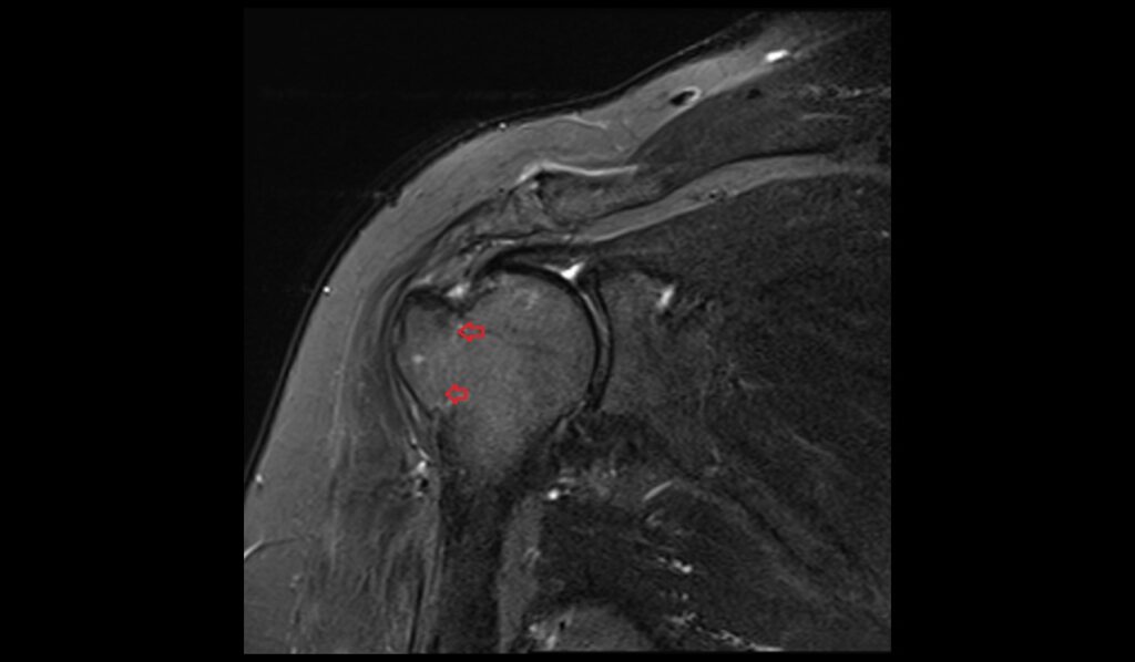
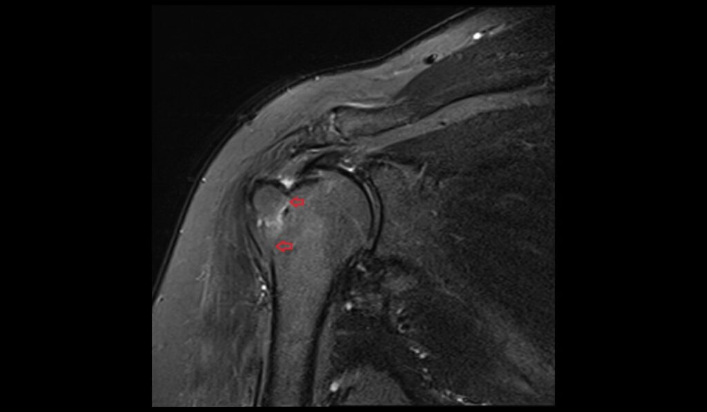
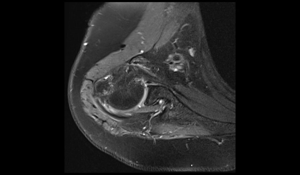
MRI PD Fat-Saturated Appearance of Greater Tuberosity Avulsion Fracture
Proton Density (PD) fat-saturated MRI sequences combine high-resolution imaging with fat suppression to enhance the visibility of fluid and edema. In the case of a greater tuberosity avulsion fracture, PD fat-saturated images typically show a hyperintense (bright) signal at the fracture site, corresponding to bone marrow edema and hemorrhage. The fat saturation technique helps to suppress the signal from the surrounding fatty tissue, allowing for a clearer view of the injury. This sequence is particularly useful for assessing associated soft tissue injuries, such as tears or inflammation in the rotator cuff tendons and other surrounding structures.
STIR coronal image shows Greater Tuberosity Avulsion Fracture
T1 TSE sagittal image shows Greater Tuberosity Avulsion Fracture
PD FS axial image shows Greater Tuberosity Avulsion Fracture

PD FS sagittal image shows Greater Tuberosity Avulsion Fracture
References
- Chillemi, C., Proietti, R., Rengo, M., Damo, M., Paolicelli, D., & Castagna, A. (2021). Fracture avulsion of the greater tuberosity: Arthroscopic transosseous augmented technique. Arthroscopy Techniques, 10(5), e1233–e1238. https://doi.org/10.1016/j.eats.2021.01.017
- Chillemi, C., Proietti, R., Rengo, M., Damo, M., Paolicelli, D., & Castagna, A. (2021). Fracture avulsion of the greater tuberosity: Arthroscopic transosseous augmented technique. Arthroscopy Techniques, 10(5), e1233-e1238.
- Longo, U.G., Corbett, S., & Ahrens, P.M. (2018). Missed fractures of the greater tuberosity. BMC Musculoskeletal Disorders, 19, Article 313. https://doi.org/10.1186/s12891-018-2262-3