MRI Gallstones
Gallstones are hard deposits that can form in the gallbladder, a small organ located under the liver. The gallbladder stores bile, a fluid produced by the liver to help digest fats. Gallstones can range in size from as small as a grain of sand to as large as a few centimeters.
Causes
Cholesterol Stones: The most common type, formed when there is too much cholesterol in the bile. Factors that may contribute include liver releasing more cholesterol than bile can dissolve, less bile salts, or poor gallbladder emptying.
Pigment Stones: These are less common and are formed when there is too much bilirubin in the bile. Conditions like liver cirrhosis, biliary tract infections, and certain blood disorders can lead to pigment stone formation.
Treatment
Observation: If gallstones are not causing symptoms, treatment may not be necessary.
Medications: Certain medications can dissolve gallstones, but this is typically reserved for those who cannot undergo surgery. These drugs can take years to be effective and the stones can recur after the treatment ends.
Surgery (Cholecystectomy): The most common and effective treatment for symptomatic gallstones is the surgical removal of the gallbladder. This is a common procedure and can often be done laparoscopically.
Endoscopic Retrograde Cholangiopancreatography (ERCP): If a stone becomes lodged in a duct, ERCP can be used to remove the stone.
MRI appearance of Gallstones
T2-Weighted MRI: On T2-weighted images, gallstones generally appear hypointense or isointense compared to the surrounding bile. The high fluid content of bile makes it appear very bright on T2, providing a good contrast with gallstones.
T2 Fat-Saturated (Fat Sat) MRI: This technique suppresses the signal from fat, making it appear dark. This is particularly useful for distinguishing between gallstones and other structures that might be high in fat.
- T1-Weighted MRI: Gallstones typically appear hypointense (dark) on T1. However, certain types of gallstones, especially those rich in cholesterol, can appear hyperintense (bright) due to their fat content.
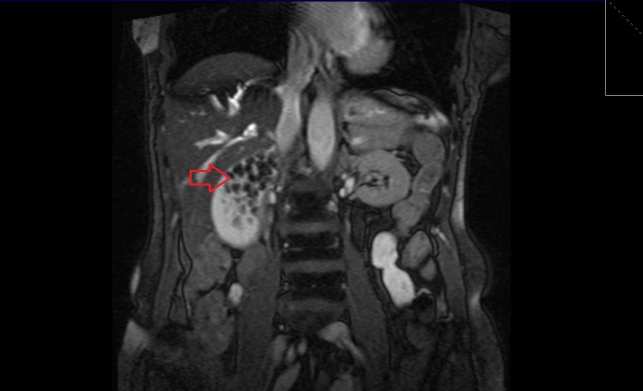
T2 TRUEFISP coronal image shows gallstones
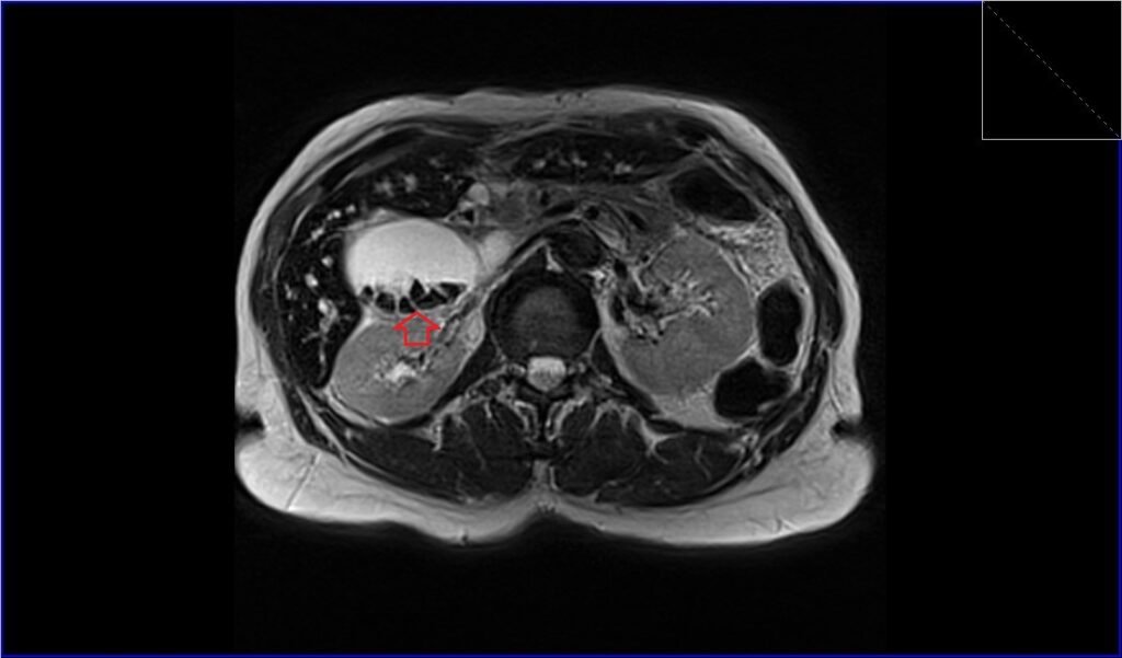
T2 TSE axial image shows gallstones
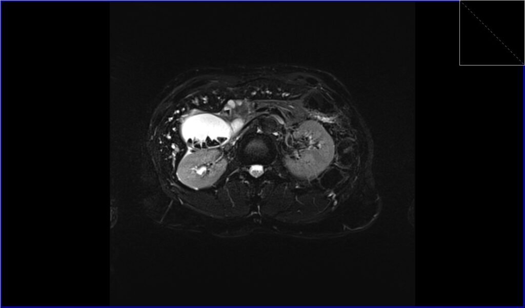
T2 Fat saturated axial image shows gallstones
References
- Ukaji, M., Ebara, M., Tsuchiya, Y., Kato, H., Fukuda, H., Sugiura, N., & Saisho, H. (2002). Diagnosis of gallstone composition in magnetic resonance imaging: in vitro analysis. European Journal of Radiology, 41(1), 49-56.
- Tsai, H.M., Lin, X.Z., Chen, C.Y., Lin, P.W., & Lin, J.C. (2004). MRI of Gallstones with Different Compositions. American Journal of Roentgenology, 182(6). https://doi.org/10.2214/ajr.182.6.1821513
- Murphy, M. C., Gibney, B., Gillespie, C., Hynes, J., & Bolster, F. (2020). Gallstones top to toe: what the radiologist needs to know. Insights into Imaging, 11, Article 13.
- Elsayes, K. M., Oliveira, E. P., Narra, V. R., EL-Merhi, F. M., & Brown, J. J. (2009). Magnetic Resonance Imaging of the Gallbladder: Spectrum of Abnormalities. Journal of Magnetic Resonance Imaging, 476-482. https://doi.org/10.1080/02841850701324102